Toggle light / dark theme

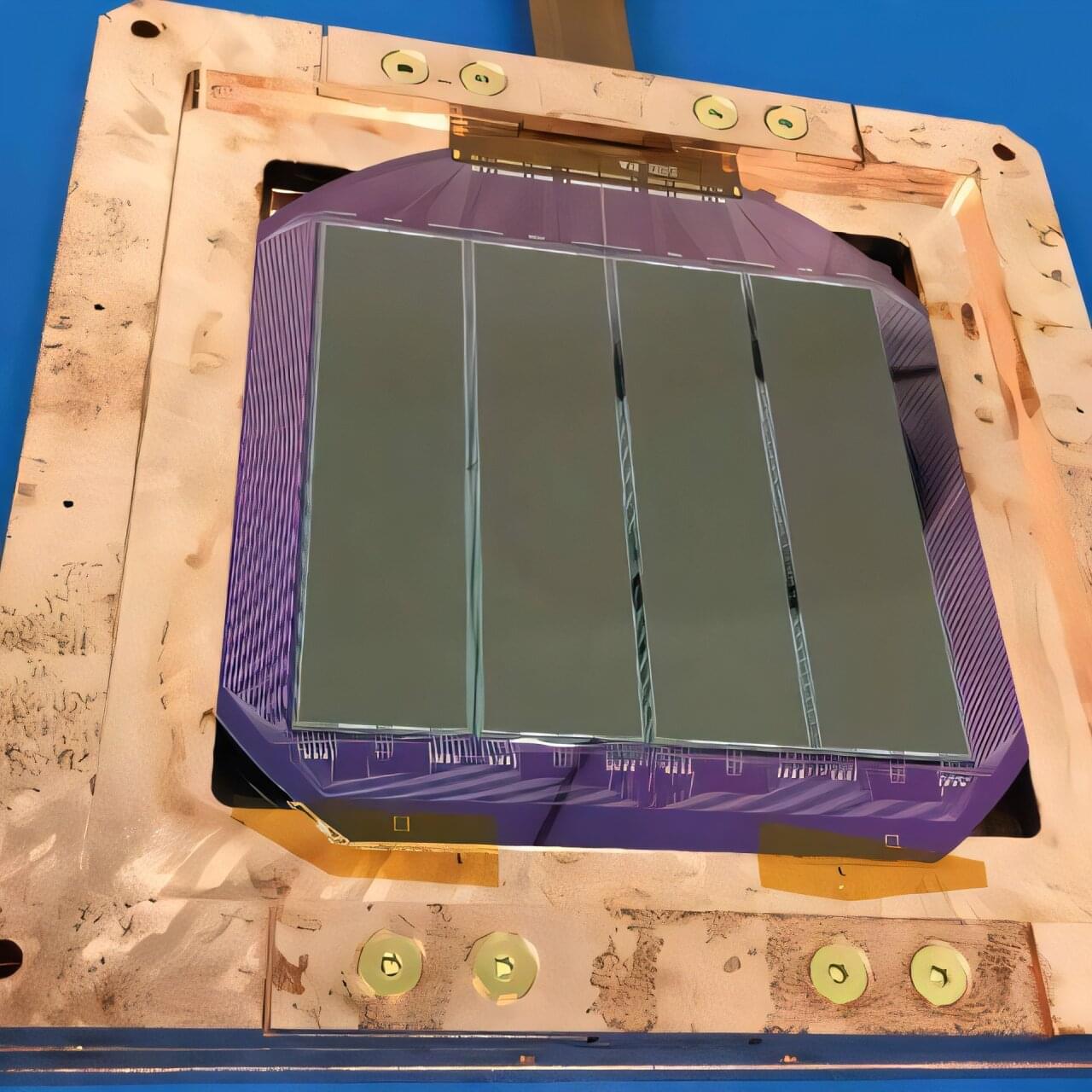
MIT physicists have taken a key step toward solving the puzzle of what leads electrons to split into fractions of themselves. Their solution sheds light on the conditions that give rise to exotic electronic states in graphene and other two-dimensional systems.

The new work is an effort to make sense of a discovery that was reported earlier this year by a different group of physicists at MIT, led by Assistant Professor Long Ju. Ju’s team found that electrons appear to exhibit “fractional charge” in pentalayer graphene — a configuration of five graphene layers that are stacked atop a similarly structured sheet of boron nitride.

Ju discovered that when he sent an electric current through the pentalayer structure, the electrons seemed to pass through as fractions of their total charge, even in the absence of a magnetic field. Scientists had already shown that electrons can split into fractions under a very strong magnetic field, in what is known as the fractional quantum Hall effect. Ju’s work was the first to find that this effect was possible in graphene without a magnetic field — which until recently was not expected to exhibit such an effect.

Researchers found that methamphetamine and PCP cause a phenomenon known as neurotransmitter switching, impairing memory. This change is reversible, pointing to potential treatments for drug-induced cognitive deficits and related neuropsychiatric conditions.

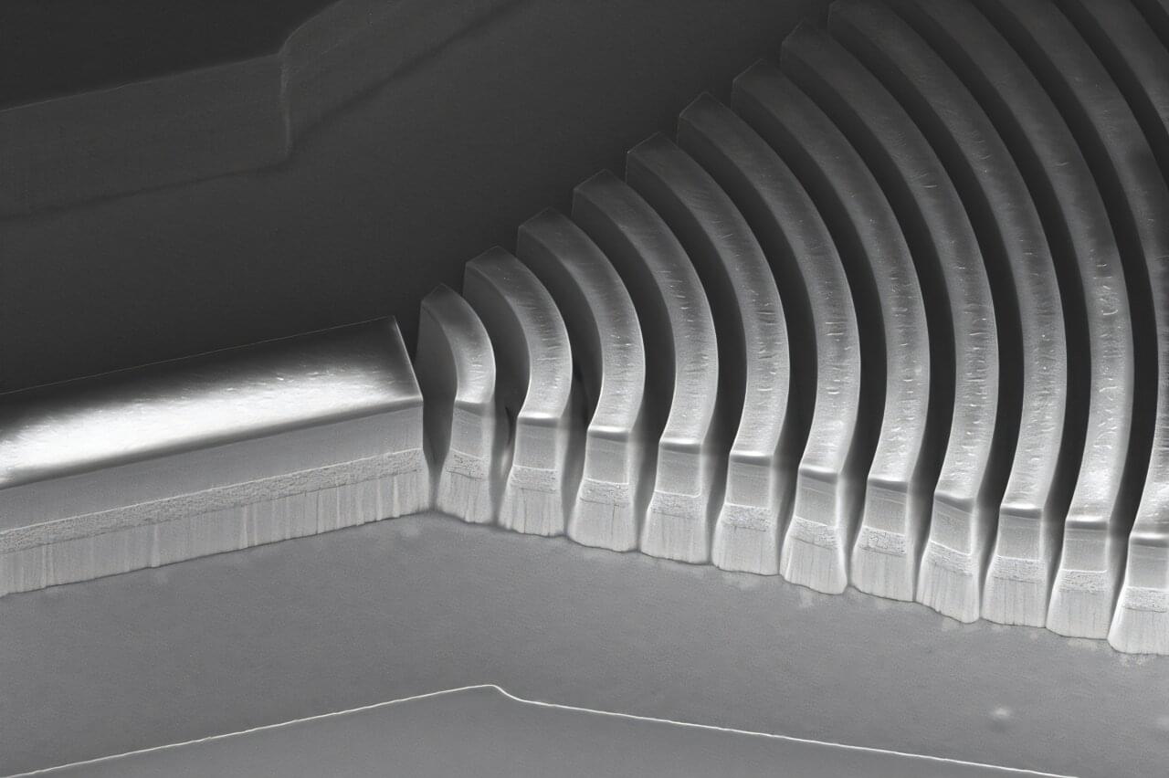
Researchers have significantly accelerated ion movement using nanotechnology, potentially improving technologies from battery charging to biosensing.

This breakthrough at Washington State University and Lawrence Berkeley National Laboratory involves creating a nanochannel lined with molecules that attract ions, allowing them to move over ten times faster than before. This development could revolutionize energy storage and help detect environmental pollutants or neurological activities.

Breaking Speed Records With Nanoscience

Stem cells are special in the way they can keep on replicating, and turn themselves into many other types of cell. Now scientists have discovered how their superpowers get a remarkable boost when they’re grown in space.

The microgravity environment increases some of the regenerative capacities of stem cells even further, researchers from the Mayo Clinic in Florida have found, based on experiments carried out on the International Space Station (ISS).

As stem cells play such a crucial role in the body’s repair process, with their ability to quickly replicate and differentiate, these findings could help in the study of disease prevention and treatment.

Acoustic Cluster Therapy (ACT®)consists of clusters of gas-filled microbubbles and oil microdroplets.


During the last decade ACT® has been evaluated preclinically in various cancer models and combined with different drugs. It was first observed that ACT was able to increase the fluorescence from a tumor when sonoenhancement was combined with fluorescent macromolecules (Wamel 2016, Figure 1). Here it was found that already one minute after sonoenhancement, fluorescence had increased in the tumor compared to a non-treated control, followed by a fluorescence uptake that remained for several hours. Subsequently, ACT has been tested therapeutically in preclinical models of prostate cancer (Wamel 2016), pancreatic cancer (Kotopoulis 2016, Ng 2022), colon cancer (Bush 2019) and breast cancer (Bush 2020). The combination of ACT and drug was significantly better than the drug alone in all these studies, with quite large numbers of complete remissions. Combining ACT with the drug nab-paclitaxel for treatment of prostate cancer resulted in complete tumor remission in all tested animals. This shows that ACT provides sonoenhancement across very different cancer types and with different types of drugs, which increases the likelihood of seeing effects also in clinical trials as the tumor models collectively represents a variety of cancer biology.

Side effects and toxicity have also been tested in various small animal models during the last decade. During treatment, no bleeding or macroscopic damage was observed, and pathological evaluation has not identified microscopic damages. ACT was extensively tested for systemic toxicities, including studies in rats and dogs where ultrasound to the heart and liver was used to activate the ACT bubbles, and no significant adverse effects have been detected.

Sonoenhancement describes the actions of ACT. This is distinct from sonoporation describing the mode of action of ultrasound with the conventional free-flowing microbubbles, which are designed for ultrasound contrast enhancement. The mode of action of ACT is that the microclusters expand and lodge in tumor capillaries. This gives multiple effects in the tumor that can be separated into primary and secondary effects. The primary effect of ACT is the oscillations inside the capillaries, which affects the vascular wall and propagate into the extravascular domain of the tumor. This is clearly different from the action achieved with conventional microbubbles. An activated ACT bubble has a volume 1,000 times that of a conventional microbubble and a large contact area with endothelial cells. An activated ACT bubble will temporarily block the capillary and oscillations will affect the entire inner surface of the vessel. The volume of the bubble and the amplitude of the oscillations results in biomechanical work that is 1,000 times greater than that of conventional microbubbles.