Toggle light / dark theme

Researchers at Karolinska Institutet have found a molecule that can both help the intestines to heal after damage and suppress tumor growth in colorectal cancer. The discovery could lead to new treatments for inflammatory bowel disease (IBD) and cancer.

The results are published in the journal Nature in a paper titled “Liver X receptor unlinks intestinal regeneration and tumorigenesis.”

Many patients with inflammatory bowel disease (IBD) such as Crohn’s disease or ulcerative colitis do not respond to available treatments, highlighting the need to identify novel therapeutic strategies. In their study, researchers propose that promoting mucosal healing through tissue regeneration could be a valid alternative to immunosuppressive drugs.

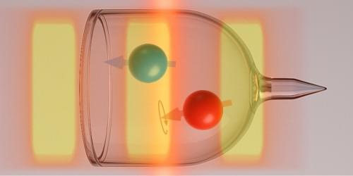
A research team led by Professor Jaedong Lee from the Department of Chemical Physics of DGIST has introduced a novel quantum state and a pioneering mechanism for extracting and controlling quantum information using exciton and Floquet states.

Collaborating with Professor Noejung Park from UNIST’s Department of Physics, the team has, for the first time, demonstrated the formation and synthesis process of exciton and Floquet states, which arise from light-matter interactions in two-dimensional semiconductors.

The study, published in Nano Letters in October, captures quantum information in real-time as it unfolds through entanglement, offering valuable insights into the exciton formation process in these materials, thereby advancing quantum information technology.

A research team led by Dr. Du Xuemin from the Shenzhen Institutes of Advanced Technology (SIAT) of the Chinese Academy of Sciences has reported a living interface with unique functionalities of durable secretion of bioactive exosomes with tunable contents and miRNA cargoes, effectively promoting neurovascular remodeling.

The study was published in Matter on Nov. 21.

Neurovascular remodeling is crucial for restoring normal functions of regenerated tissues or engineered organs, which requires multi-target and phase-specific paracrine regulation. However, existing strategies still cannot mimic such dynamic and complicated paracrine regulation effects in the native physiological processes, hindering synergistic neurovascular remodeling.

Communication and coordination among different cells are fundamental aspects that regulate many functions in our body. This process, known as paracrine signaling, involves the release of signaling molecules by a cell into its extracellular matrix (ECM) or surroundings to communicate changes in its cellular processes or the local environment. These signaling molecules are then detected by neighboring cells, leading to various cellular responses.

For instance, during cell/tissue injury, the paracrine signaling process releases that signal nearby stem cells to assist in tissue repair in the form of scar tissue formation or blood clotting. Similar processes occur in the regulation of other vital functions, such as digestion, respiration, and reproduction. Additionally, paracrine signals influence the expression and activity of enzymes involved in drug metabolism and play a role in drug–drug interactions.

The signaling molecules, which may contain proteins and , are transported within tiny vesicles called exosomes. These vesicles serve as valuable biomarkers for various diseases and can even be engineered to carry drugs, making them a highly effective targeted drug delivery system. Notably, the hormone oxytocin and the neurotransmitter dopamine are paracrine messengers.

Researchers have used 3D cell culture models in the past decade to translate molecular targets during drug discovery processes to thereby transition from an existing predominantly 2D culture environment. In a new report now published in Science Advances, Charalampos Pitsalidis and a research team in physics and chemical engineering at the University of Science and Technology in Abu Dhabi, UAE and the University of Cambridge describe a multi-well plate bioelectronic platform named the e-transmembrane to support and monitor complex 3D cell architectures.

The team microengineered the scaffolds using poly(3,4-ethylenedioxythiophene polystyrene sulfonate to function as separating membranes to isolate cell cultures and achieve real-time in situ recordings of cell growth and function. The to volume ratio allowed them to generate deep stratified tissues in a porous architecture. The platform is applicable as a universal resource for biologists to conduct next-generation high-throughput drug screening assays.

Insecticides have been used for centuries to counteract widespread pest damage to valuable food crops. Eventually, over time, beetles, moths, flies and other insects develop genetic mutations that render the insecticide chemicals ineffective.

Escalating resistance by these mutants forces farmers and vector control specialists to ramp up use of poisonous compounds at increasing frequencies and concentrations, posing risks to human health and damage to the environment since most insecticides kill both ecologically important insects as well as pests.

To help counter these problems, researchers recently developed powerful technologies that genetically remove insecticide-resistant variant genes and replace them with genes that are susceptible to pesticides. These gene-drive technologies, based on CRISPR gene editing, have the potential to protect valuable crops and vastly reduce the amount of chemical pesticides required to eliminate pests.

Scientists have discovered that tRNAs can determine how long mRNAs exist in a cell, causing some messages to be stabilized and translated into more protein, while directing others to be degraded and limiting how much protein can be made. They published their report in Science.

The messenger RNA (mRNA)-based vaccines developed to fight the virus SARS-CoV-2 saved lives and made the nucleic acid a household name during the COVID-19 pandemic. Suddenly, everyone knew a little bit more about the molecule that helps convert genetic information into proteins.

But in addition to determining which proteins are made, mRNAs can also specify how much protein is produced.

A multi-institutional team of researchers, led by Georgia Tech’s Francesca Storici, has discovered a previously unknown role for RNA. Their insights could lead to improved treatments for diseases like cancer and neurodegenerative disorders while changing our understanding of genetic health and evolution.

A new “toolkit” to repair damaged DNA that can lead to aging, cancer and motor neuron disease (MND) has been discovered by scientists at the Universities of Sheffield and Oxford.

Published in Nature Communications, the research shows that a protein called TEX264, together with other enzymes, is able to recognize and “eat” toxic proteins that can stick to DNA and cause it to become damaged. An accumulation of broken, damaged DNA can cause cellular aging, cancer and neurological diseases such as MND.

Until now, ways of repairing this sort of DNA damage have been poorly understood, but scientists hope to exploit this novel repair toolkit of proteins to protect us from aging, cancer and neurological disease.

Junk DNA could unlock new treatments for neurological disorders as scientists discover its breaks and repairs affect our protection against neurological disease.

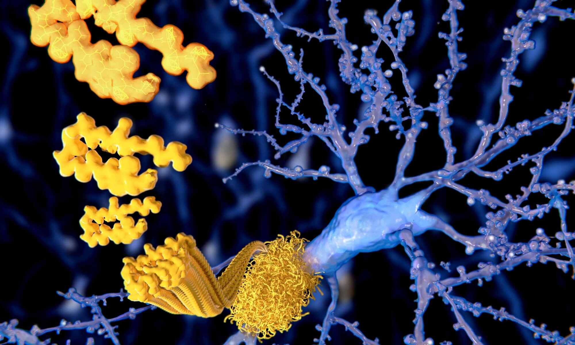
The research from the University of Sheffield’s Neuroscience Institute and Healthy Lifespan Institute gives important new insights into so-called junk DNA and how it impacts on neurological disorders such as Motor Neuron Disease (MND) and Alzheimer’s.

Until now, the repair of junk DNA, which makes up 98% of DNA, has been largely overlooked by scientists, but the new study published in Nature found it is much more vulnerable to breaks from oxidative genomic damage than previously thought. This has vital implications on the development of neurological disorders.