Researchers developed CrystaLLM, an AI model predicting crystal structures without traditional physics-based simulations. By analyzing millions of existing structures, it predicts realistic arrangements of atoms, even for unfamiliar materials.
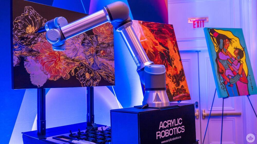
Montreal-based Acrylic Robotics is utilizing a robot’s arm to paint fine art on canvas using AI software that emulates the actual painter’s brush strokes.
The startup showcased its technology with demos at AWS re: Invent, Amazon’s cloud service conference held in Las Vegas, where an AI robot dutifully worked on a painting, or “Auragraph,” live. Holding a brush, it would carefully dip into the different pools of acrylic paint below and then position the brush to apply a stroke at just the right spot.
In a sense, it felt a little like watching an automated assembly, only in a very obvious artistic context. Acrylic Robotics is trying to meld the worlds of artificial intelligence, engineering, robotics and art into a practical form of production. The idea is not to just produce replicas of an artist’s pieces, but to also bring digital creations to canvas without resorting to simple prints. At the same time, the company’s ethical approach is to ensure artists get paid for what they create.
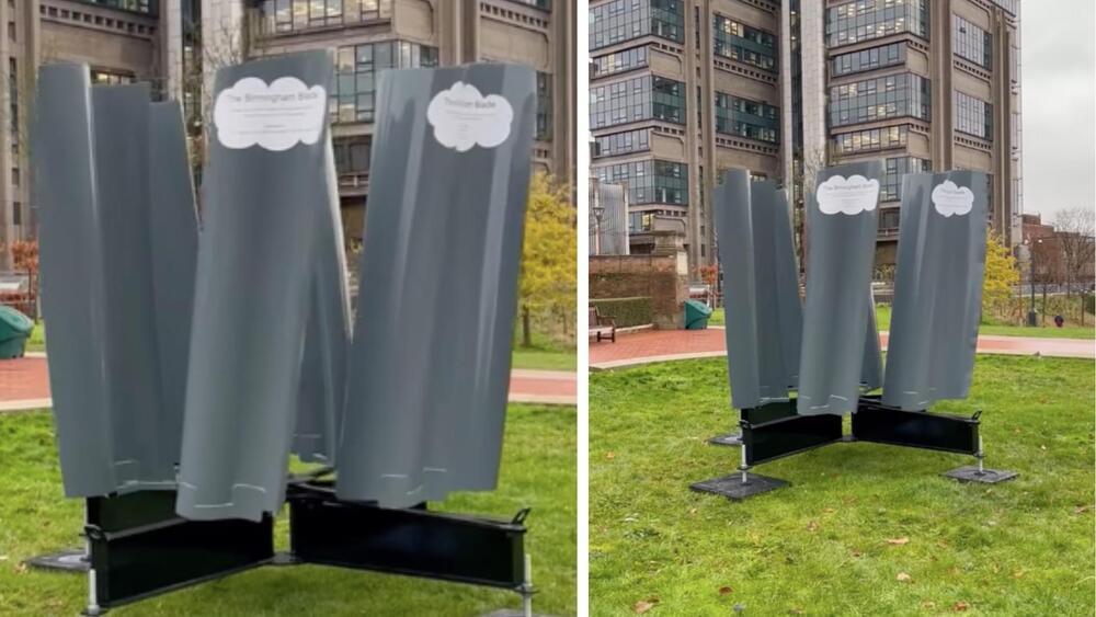
The world’s first urban wind turbine designed by AI has been unveiled in the UK.
Called the Birmingham Blade, the turbine is jointly developed by AI design specialists EvoPhase and precision metal fabricators KwikFab. The turbine is also tailored to the unique wind conditions of a specific geographic area.
EvoPhase claimed that it used its AI-driven design process to generate and test designs for their efficiency at wind speeds found in Birmingham, which, at 3.6 meters/second are substantially lower than the 10 meters/second rating for most turbines.

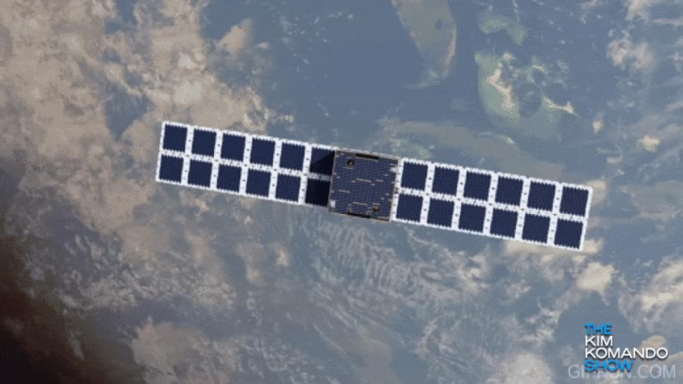
An international team of scientists using observations from NASA-German satellites found evidence that Earth’s total amount of freshwater dropped abruptly starting in May 2014 and has remained low ever since. Reporting in Surveys in Geophysics, the researchers suggested the shift could indicate Earth’s continents have entered a persistently drier phase.
From 2015 through 2023, satellite measurements showed that the average amount of freshwater stored on land—that includes liquid surface water like lakes and rivers, plus water in aquifers underground—was 290 cubic miles (1,200 cubic km) lower than the average levels from 2002 through 2014, said Matthew Rodell, one of the study authors and a hydrologist at NASA’s Goddard Space Flight Center in Greenbelt, Maryland. “That’s two and a half times the volume of Lake Erie lost.”
During times of drought, along with the modern expansion of irrigated agriculture, farms and cities must rely more heavily on groundwater, which can lead to a cycle of declining underground water supplies: freshwater supplies become depleted, rain and snow fail to replenish them, and more groundwater is pumped.

Researchers at The Hospital for Sick Children (SickKids) have uncovered that stress changes how our brain encodes and retrieves aversive memories, and discovered a promising new way to restore appropriate memory specificity in people with post-traumatic stress disorder (PTSD).
If you stumble during a presentation, you might feel stressed the next time you have to present because your brain associates your next presentation with that one poor and aversive experience. This type of stress is tied to one memory.
But stress from traumatic events like violence or generalized anxiety disorder can spread far beyond the original event, known as stress-induced aversive memory generalization, where fireworks or car backfires can trigger seemingly unrelated fearful memories and derail your entire day. In the case of PTSD, it can cause much greater negative consequences.
I have my own introduction to quantum mechanics course that you can check out on Brilliant! First 30 days are free and 20% off the annual premium subscription when you use our link ➜ https://brilliant.org/sabine.
“New physics” is a catch-all term for fundamentally new discoveries in physics (such as dark matter, quantum gravity, or a theory of everything) which push the boundaries of how we understand our reality. How could new discoveries in these areas of research affect our lives? Let’s take a look at what knowledge and practical use we could potentially gain.
🤓 Check out my new quiz app ➜ http://quizwithit.com/
💌 Support me on Donorbox ➜ https://donorbox.org/swtg.
📝 Transcripts and written news on Substack ➜ https://sciencewtg.substack.com/
👉 Transcript with links to references on Patreon ➜ / sabine.
📩 Free weekly science newsletter ➜ https://sabinehossenfelder.com/newsle…
👂 Audio only podcast ➜ https://open.spotify.com/show/0MkNfXl…
🔗 Join this channel to get access to perks ➜
/ @sabinehossenfelder.
🖼️ On instagram ➜ / sciencewtg.
#science #physics
Subboor Ahmad is a Muslim evolution critic who likes to quote academics to support his case. One in particular is Elliott Sober, whom Subboor described as \.
Sean Michael Carroll (born 5 October 1966) is a cosmologist and Physics professor specializing in dark energy and general relativity. He is a research professor in the Department of Physics at the California Institute of Technology. He has been a contributor to the physics blog Cosmic Variance, and has published in scientific journals and magazines such as Nature, Seed, Sky \& Telescope, and New Scientist.
https://en.wikipedia.org/wiki/Sean_M…
Other videos related to challenging or debunking the fine tuning argument
• Video.
A Rebuttal to the Fine-Tuning Argument.
• A Rebuttal to the Fine-Tuning Argument.