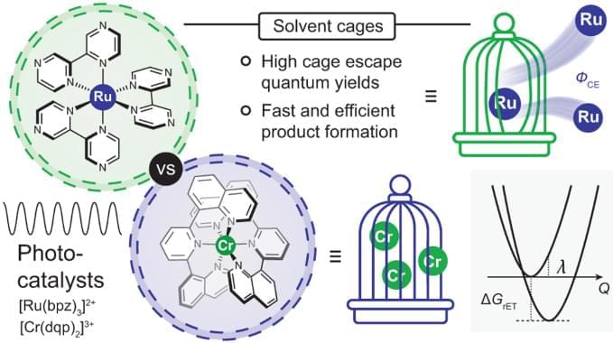
Toggle light / dark theme

Researchers at Weill Cornell Medicine have uncovered a novel route to stimulate the growth of healthy insulin-producing pancreatic beta cells in a preclinical model of diabetes. The findings hold promise for future therapeutics that will improve the lives of individuals with type 2 diabetes—a condition that affects more than half a billion people worldwide.

This study, published in the Journal of Clinical Investigation on Sept. 15, demonstrated that activating a pathway to promote not only expanded the population of insulin-producing cells, but surprisingly, it also enhanced the cells’ function.

“That’s reassuring because there is a long-standing belief in the field that proliferation can lead to ‘de-differentiation’ and a loss of cell function,” said study senior author Dr. Laura Alonso, chief of the division of endocrinology, and metabolism, director of the Weill Center for Metabolic Health, and the E. Hugh Luckey Distinguished Professor in Medicine at Weill Cornell Medicine. “Our result flies in the face of that dogma and suggests if we can find a way to trigger replication of the in the body, we won’t impair their ability to produce and secrete insulin.”

Thyrotoxicosis was associated with 39% higher risk for cognitive disorders.

Thyrotoxicosis, defined as a low level of serum thyroid-stimulating hormone (TSH), can result from either a primary thyroid disorder (endogenous) or overtreatment of hypothyroidism (exogenous). Evidence suggests that thyrotoxicosis is a risk factor for dementia. In this U.S. longitudinal cohort study, researchers used data from electronic health records for 66,000 people (median age, 68) without low TSH levels or cognitive disorders at baseline and evaluated whether development of thyrotoxicosis was associated with excess risk for cognitive disorders.

During the study period (2014 to 2023), 2,700 patients had low TSH levels (60% exogenous), and 4,800 patients received diagnoses of cognitive disorders. The incidence of cognitive disorders among patients with and without thyrotoxicosis were 11% and 6% at age 75, and 34% and 26% at age 85. Adjusted for multiple variables, all-cause thyrotoxicosis was associated with a significant 39% excess risk for cognitive disorders. Exogenous thyrotoxicosis — and in particular, severe exogenous thyrotoxicosis (TSH 0.1 mIU/L) — were associated most strongly with excess risk for cognitive disorders.

“You can see a very strong modulation, which is always there. As the modulation gets to be more profound, it eventually flattens out, and that’s when the brain reaches the deeper state,” Brown says.

When the amount of drug was reduced, the amplitude of the alpha waves began to increase again.

The researchers also found a distinctive pattern in the slow and delta waves seen in the patients’ EEG readings. Slow and delta oscillations are the slowest brain waves, and as the amount of drug was increased, the frequency of these waves became slower and slower, reflecting a decrease in brain activity.

Memory, a fundamental tool for our survival, is closely linked with how we encode, recall, and respond to external stimuli. Over the past decade, extensive research has focused on memory-encoding cells, known as engram cells, and their synaptic connections. Most of this research has centered on excitatory neurons and the neurotransmitter glutamate, emphasizing their interaction between specific brain regions.

To expand the understanding of memory, a research team led by KAANG Bong-Kiun (Seoul National University, Institute of Basic Science) developed a technology called LCD-eGRASP (local circuit dual-eGRASP) that can label synapses of neural circuits within a specific brain region. The team applied this new technology to identify the local synaptic connections between inhibitory interneurons and engram cells, shedding light on the role of inhibitory interneurons in memory expression.

The researchers targeted basolateral amygdala (BLA), an evolutionarily well-preserved brain region in vertebrates known for controlling positive and negative emotions in animals, especially . When a fear-related event occurs, neurons activated during that specific time point become engram cells, encoding the .

In a recent study published in Frontiers in Psychology, researchers evaluate the association between paternal mental health and a child’s development during middle childhood.

Study: Longitudinal associations between paternal mental health and child behavior and cognition in middle childhood. Image Credit: PeopleImages.com — Yuri A/Shutterstock.com.

The human brain is three times bigger than a chimp’s and more spherical than a Neanderthal’s. Within a maze of bumps and grooves, neurons converse in distinct patterns that give humans unique cognitive abilities.

Scientists haven’t fully deciphered those patterns. But researchers at UT Southwestern Medical Center are determined to solve the molecular mystery of what makes us .

In a study published in the journal Nature, they compared brain cell types and activities among humans, chimpanzees and rhesus monkeys. Human brains had more of a kind of cell that may help them adapt based on new experience and heal from injury. Certain human neurons also had more of a gene that affects language development.