The optical crystal can survive extreme temperatures and cosmic radiation, is nigh-impervious to physical impact, resists most chemicals, and can endure for billions of years.
Get the latest international news and world events from around the world.
Revolution in medicine: A molecule produced by gut bacteria causes atherosclerosis, responsible for millions of deaths
The Argentine microbiologist Federico Rey and Indian pathologist Vaibhav Vemuganti applaud the “exciting opportunities” that the new study opens for the prevention and treatment of cardiovascular disease. In a commentary also published Wednesday in Nature, the two experts emphasize that exposure to imidazole propionate worsens plaque formation in the arteries of mice. “This effect occurs independently of changes in cholesterol levels, a surprising result given the central role of cholesterol in the development of atherosclerosis,” note the two specialists, from the University of Wisconsin-Madison. “This discovery offers an interesting clue about a possible new factor involved in the origin of atherosclerosis. This is very relevant because, although lowering cholesterol — through drugs called statins, for example — can effectively reduce the risk of cardiovascular disease, a considerable proportion of people still experience adverse cardiovascular events, such as myocardial infarctions or strokes,” they warn. The CNIC itself said in a statement that the new study “could revolutionize” the diagnosis and treatment of atherosclerosis.
Sancho stresses that the work has been made possible thanks to the collaboration of thousands of volunteer employees of Banco Santander in Madrid, but also thanks to grants of €1 million from the “la Caixa” Foundation, €150,000 from the European Research Council and €100,000 from the State Research Agency.
The discovery of the decisive effect of imidazole propionate on atherosclerosis takes place against a backdrop in which the scientific community is revealing the unknown role of intestinal microbes in some human diseases. The biotechnologist Cayetano Pleguezuelos and his colleagues at the Hubrecht Institute (The Netherlands) demonstrated in February 2020 that a strain of the bacterium Escherichia coli produces a toxic molecule, called colibactin, which damages the DNA of human cells and causes malignant tumors.
Entangled Atomic Clock Experiment Could Finally Provide Hints At A Theory Of Everything
A new experiment involving a network of entangled atomic clocks could finally help us test how quantum mechanics fits with general relativity.
Quantum mechanics is our best understanding of the universe at atomic and subatomic scales. With it, we have revolutionized our understanding of physics on teeny tiny scales. General relativity – first outlined by Albert Einstein in 1915 – meanwhile, is our best understanding of gravity. According to the theory, which has so far passed every test we have thrown at it, gravity is not a force but the result of the curvature of spacetime around matter.
Why AI chatbots lie to us
A few weeks ago, a colleague of mine needed to collect and format some data from a website, and he asked the latest version of Anthropic’s generative AI system, Claude, for help. Claude cheerfully agreed to perform the task, generated a computer program to download the data, and handed over perfectly formatted results. The only problem? My colleague immediately noticed that the data Claude delivered was entirely fabricated.
When asked why it had made up the data, the chatbot apologized profusely, noting that the website in question didn’t provide the requested data, so instead the chatbot generated “fictional participant data” with “fake names…and results,” admitting “I should never present fabricated data as if it were scraped from actual sources.”
I have encountered similar examples of gaslighting by AI chatbots. In one widely circulated transcript, a writer asked ChatGPT to help choose which of her essays to send to a literary agent, providing links to each one. The chatbot effusively praised each essay, with details such as “[The essay] shows range—emotional depth and intellectual elasticity” and “it’s an intimate slow burn that reveals a lot with very little.” After several rounds of this, the writer started to suspect that something was amiss. The praise was effusive, but rather generic. She asked “Wait, are you actually reading these?” ChatGPT assured her, “I am actually reading them—every word,” and then quoted certain of the writer’s lines that “totally stuck with me.” But those lines didn’t actually appear in any of the essays. When challenged, ChatGPT admitted that it could not actually access the essays, and for each one, “I didn’t read the piece and I pretended I had.”
A new analysis of the neurocranium and mandible of the Skhūl I child: Taxonomic conclusions and cultural implications
0,
In a study published last week in the journal L’Anthropologie, researchers re-analyzed fragments of Skhūl I, the name for remains belonging to a likely female child between the ages of 3 and 5. While the individual is currently recognized as an anatomically modern human, Homo sapiens, its classification remains contentious, given that it has some Neanderthal-like features. Now, the new study suggests the child might have been a hybrid—and potentially had one Homo sapiens parent and one Neanderthal parent.
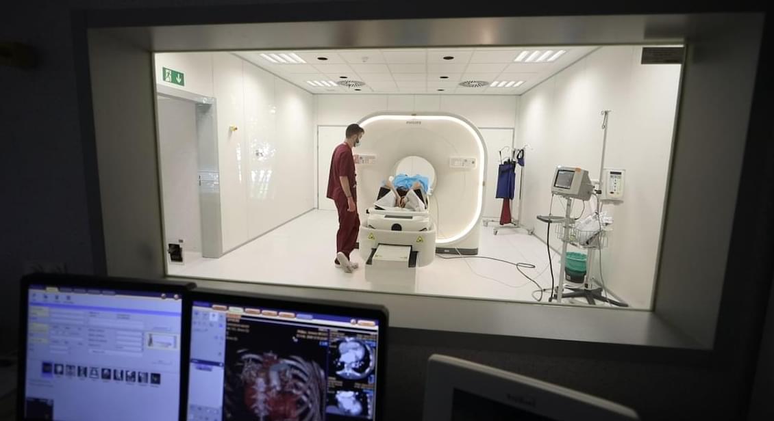
To reach this conclusion, the team conducted CT scans of the child’s neurocranium—the part of the skull that protects the brain—and jaw. They compared the resulting 3D models to remains of other Homo sapiens and Neanderthal children. In short, they found the neurocranium to be more similar to that of a modern human, while the jaw was more akin to a Neanderthal’s.
“The combination of features seen in Skhūl I may suggest that the child is a hybrid,” the researchers write in the study. “In the Middle Pleistocene, the Levant was the crossroad of gene flows between Indigenous lineages and other taxa from Africa and Eurasia, which is likely the explanation for Skhūl I anthropological.”
Their results align with genetic evidence indicating that modern humans and Neanderthals didn’t just cross paths—they interbred for thousands of years. In fact, some research has suggested Homo sapiens drove Neanderthals to extinction not with violence, but by absorbing them into their population through interbreeding. Regardless of the reason for Neanderthals’ demise, many humans have Neanderthal DNA today.
nouvelle analyse du neurocr ne et de la mandibule de l’enfant Skhūl I : conclusions taxonomiques et implications culturelles.
Eco-friendly 3D-printed house uses soil, not cement — building still scores top earthquake resistance rating
The material reminds us of wattle and daub, a home building material harking back 6,000 years.
104 Quasi-Stellar Objects Studied Using Telescope Reveal Stunning Details
A group of astronomers, led by researchers at Rhodes University, has analyzed 104 quasars through the MIGHTEE survey, a large-scale project using South Africa’s MeerKAT radio telescope.