Believe it or not I’m posting new music on Soundcloud!https://soundcloud.com/dilliot2kListen to my podcast!https://anchor.fm/dilliot2kFollow my weight loss j…
Get the latest international news and world events from around the world.


World’s First Universal Intelligent Human “Tong Tong” 2.0 Released, Expected to Have the Intelligence Level of 6-Year-Old Children Within the Year — ai, artificial intelligence,1ai.net
March 30, 2012 — At yesterday’s 2025 Zhongguancun Forum At the annual meeting, the Beijing General Artificial Intelligence Research Institute launched theThe world’s first Universal Intelligent Man“complete” 2.0 officially released.
“Tom-Tom” is positioned as a virtual human with autonomous learning, cognitive and decision-making capabilities. Expected to have the intelligence of a 6 year old within this year..

Plants captured on video communicating with each other for the first time ever
This study builds on observations first made in 1983, which sparked debates and further research into plant communication.
Over the years, scientists have uncovered various ways plants interact, from chemical signals to underground networks formed by fungi.
“We have finally unveiled the intricate story of when, where, and how plants respond to airborne ‘warning messages’ from their threatened neighbors,” Dr. Toyota emphasized.

Artificial neurons organize themselves
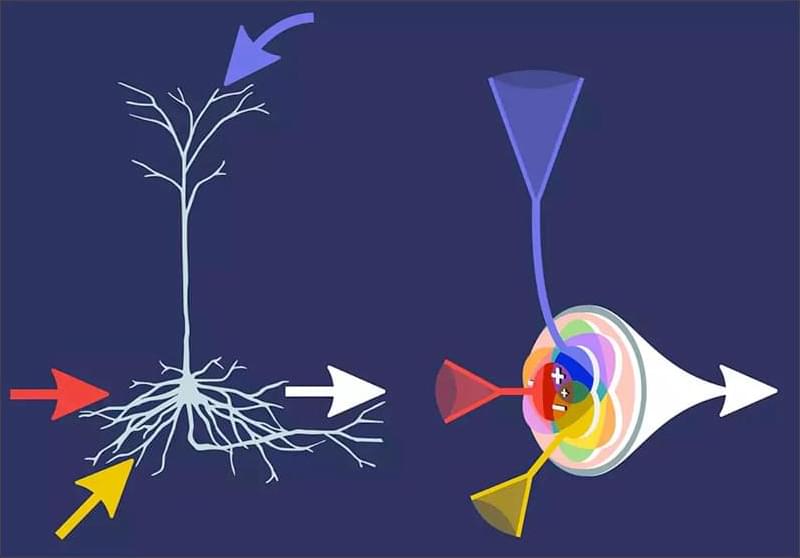
Novel artificial neurons learn independently and are more strongly modeled on their biological counterparts. A team of researchers from the Göttingen Campus Institute for Dynamics of Biological Networks (CIDBN) at the University of Göttingen and the Max Planck Institute for Dynamics and Self-Organization (MPI-DS) has programmed these infomorphic neurons and constructed artificial neural networks from them. The special feature is that the individual artificial neurons learn in a self-organized way and draw the necessary information from their immediate environment in the network.
The results were published in PNAS (“A general framework for interpretable neural learning based on local information-theoretic goal functions”).
Both, human brain and modern artificial neural networks are extremely powerful. At the lowest level, the neurons work together as rather simple computing units. An artificial neural network typically consists of several layers composed of individual neurons. An input signal passes through these layers and is processed by artificial neurons in order to extract relevant information. However, conventional artificial neurons differ significantly from their biological models in the way they learn.