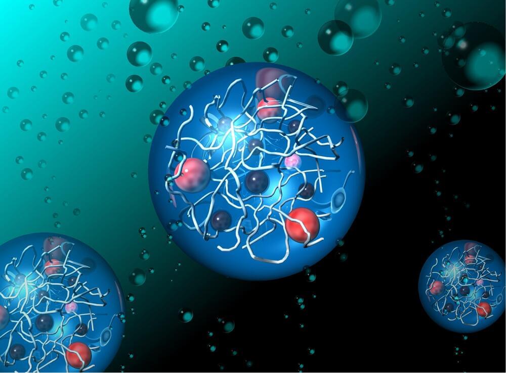
As the efficiency of non-viral delivery methods improves, they have the potential to make cell and gene therapy development faster and more cost-effective, as well as mitigating adverse effects.
Salk Institute researchers, as part of a larger collaboration with research teams around the world, analyzed more than half a million brain cells from three human brains to assemble an atlas of hundreds of cell types that make up a human brain in unprecedented detail.
The research, published in a special issue of the journal Science on October 13, 2023, is the first time that techniques to identify brain cell subtypes originally developed and applied in mice have been applied to human brains.
“These papers represent the first tests of whether these approaches can work in human brain samples, and we were excited at just how well they translated,” says Professor Joseph Ecker, director of Salk’s Genomic Analysis Laboratory and a Howard Hughes Medical Institute investigator. “This is really the beginning of a new era in brain science, where we will be able to better understand how brains develop, age, and are affected by disease.”
Whether in the brain or in the muscles, wherever there are nerve cells, there are synapses. These contact points between neurons form the basis for the transmission of excitation, the communication between neurons. As in any communication process, there is a sender and a receiver: Nerve cell processes called axons generate and transmit electrical signals thereby acting as signal senders.
Synapses are points of contact between axonal nerve terminals (the pre-synapse) and post-synaptic neurons. At these synapses, the electrical impulse is converted into chemical messengers that are received and sensed by the post-synapses of the neighboring neuron. The messengers are released from special membrane sacs called synaptic vesicles.
As well as transmitting information, synapses can also store information. While the structure and function of synapses are comparably well understood, little is known about how they are formed.
A quarter-century ago, researchers discovered that adults, not just developing infants, can generate new brain cells, a process called neurogenesis. But it’s still not clear what role these new neurons play in health or disease.
In a new mouse study, Columbia University researchers found that neurogenesis in adults is critical for maintaining brain circuits that support working memory across the lifespan and chronic loss of adult neurogenesis causes progressive memory loss, like that seen in age-related cognitive decline and Alzheimer’s disease in humans.
The study, “Adult-born neurons maintain hippocampal cholinergic inputs and support working memory during aging,” was published in July in the journal Molecular Psychiatry.
A study by researchers in the laboratory of Dr. Huda Zoghbi, distinguished service professor at Baylor College of Medicine and director of the Jan and Dan Duncan Neurological Research Institute (Duncan NRI) at Texas Children’s Hospital, has discovered that diminished memory recall in Rett syndrome mice can be restored by activating specific inhibitory cells in the hippocampus. The findings are published in the current edition of Neuron.
Rett syndrome is a neurodevelopmental disorder characterized by loss of acquired cognitive, motor, language and social skills after the first year of life as well as profound learning and memory impairments. In particular, contextual memories, those that encode an event and the circumstances in which the event was experienced, are diminished in mouse models of Rett syndrome. Previous research has suggested that diminished contextual memories result from disruptions in the finely tuned balance between excitatory and inhibitory synaptic inputs that constantly bombard hippocampal neurons.
Zoghbi’s team hypothesized that disruptions in this balance may alter the size and composition of ensembles of hippocampal neurons needed to encode a contextual memory. Using a miniature microscope, they directly monitored these ensembles as mice recalled a fearful experience. They found that Rett mice have larger and more correlated ensembles of neurons than wild-type mice, suggesting that hippocampal pyramidal neurons are not receiving enough inhibition in Rett mice.
Developing brains become shaped by the sights, sounds, and experiences of early life. The brain’s circuits grow more stable as we age. However, some experiences later in life open up opportunities for these circuits to be rapidly rewired. New research from Cold Spring Harbor Laboratory Associate Professor Stephen Shea helps explain how the brain adapts during a critical period of adulthood: the time when new mothers learn to care for their young.
Shea’s work in mice shows how this learning process is disrupted when a small set of neurons lack a protein called MECP2. In humans, MECP2 dysfunction causes the rare neurodevelopmental disorder Rett syndrome. Shea’s findings could point researchers toward the brain circuits involved in Rett syndrome and potential treatment strategies. His research could also have implications for more common neurological conditions.
Shea explains, “It’s not lost on us that Rett syndrome patients have difficulty interpreting and producing language. Difficulties with communicating are widespread in autism spectrum disorders. One of the reasons we study Rett syndrome is that this may be a valuable model for other forms of autism.”
Central learning and memory hubs change in response to sex hormones. A new study in Nature Mental Health by Rachel Zsido and Julia Sacher of the Max Planck Institute for Human Cognitive and Brain Sciences and the University Clinic in Leipzig, Germany, links rhythmic oscillations in ovarian hormone levels in women during the menstrual cycle to changes in brain structure.
Ovarian hormones have significant effects on the brain, and early menopause may be associated with an increased risk of accelerated brain aging and dementia later in life. However, the effects of ovarian hormone fluctuations on brain structure earlier in life are less defined. In their current study, Zsido and Sacher show that fluctuations in ovarian hormones affect structural plasticity in key brain regions during the reproductive years.
To do this, the scientists collected blood samples from 27 female study participants, used ultrasound to track follicle growth in the ovaries to pinpoint ovulation timing, and utilized ultra-high field 7 Tesla MRI to zoom into subregions of the medial temporal lobe and hippocampus. That’s because these regions are dense with sex hormone receptors and are critical for cognitive function, such as episodic memory.
Scroll Prize.
But they were so delicate and prone to turn ash if mishandled that they couldn’t be read. Previous scientists studying the scrolls could make out some Greek letters and symbols and even trained a machine-learning algorithm to read the blank ink of Herculaneum.
Year 2022 face_with_colon_three
When the team fired their ultra-fast laser at a graphene wire strung between two gold electrodes, it produced two different kinds of currents. Some of the electrons excited by the light continued moving in a particular direction once the light was switched off, while others were transient and were only in motion while the light was on. The researchers found that they could control the type of current created by altering the shape of their laser pulses, which was then used as the basis of their logic gate.
Logic gates work by taking two inputs—either 1 or 0—processing them, and providing a single output. The exact processing rules depend on the kind of logic gate implementing them, but for example, an AND gate only outputs a 1 if both its inputs are 1, otherwise it outputs a 0.
In the researchers’ new scheme, two synchronized lasers are used to create bursts of either the transient or permanent currents, which act as the inputs to the logic gate. These currents can either add up or cancel each other to provide the equivalent of a 1 or 0 as an output.
VIEW NOW: https://ow.ly/GMX050PMTMH
In this webinar, three experts will discuss how Precision NanoSystems’ modular microfluidic platform technologies and analytics can help scientists successfully design, develop, test, and scale-up promising mRNA-LNP vaccines and therapeutics from concept to clinic. Don’t miss this webinar, now available on demand.
Nucleic acids (e.g., siRNA, mRNA and saRNA) can be designed and formulated to silence, express, and edit specific genes providing a flexible and powerful approach to preventing and treating diseases. The recent commercialization and widespread distribution of COVID-19 mRNA vaccines has exemplified the massive potential of this new class of genomic medicines and vaccines to effectively thwart emerging viral threats and treat a wide range of challenging diseases. Part of developing a successful mRNA therapeutic or vaccine is choosing a delivery mechanism that protects the nucleic acids on the way to their target tissue. Encapsulating mRNA in lipid nanoparticles has proven to be one of the best vehicles for overcoming extracellular and intracellular barriers and safely delivering the treatment. Several mRNA-LNP formulations that target things like viral infections and cancers are being evaluated clinically.
In this webinar, three experts will discuss how Precision Nanosystems’ modular microfluidic platform technologies and analytics can help scientists successfully design, develop, test, and scale-up promising mRNA-LNP vaccines and therapeutics from concept to clinic. They will provide an overview of Precision NanoSystem’s Biopharma Services, and share examples from internal R&D work that demonstrate the versatility of the genetic medicine toolbox for rapidly developing RNA-LNP vaccines. You’ll also learn about Precision NanoSystem’s BioAssay services and the capabilities that are available to facilitate and accelerate drug development projects.