Toggle light / dark theme

Get the latest international news and world events from around the world.

Log in for authorized contributors

JWST may have found the Universe’s first stars powered by dark matter

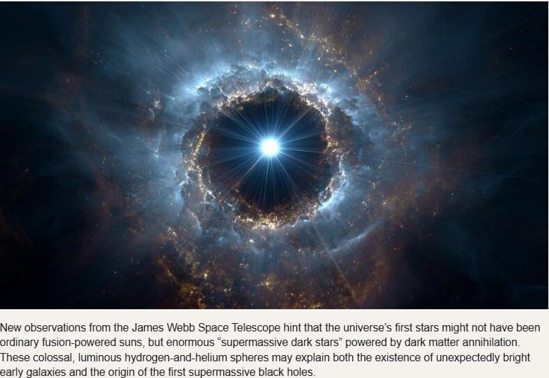
New observations from the James Webb Space Telescope hint that the universe’s first stars might not have been ordinary fusion-powered suns, but enormous “supermassive dark stars” powered by dark matter annihilation. These colossal, luminous hydrogen-and-helium spheres may explain both the existence of unexpectedly bright early galaxies and the origin of the first supermassive black holes.

In the early universe, a few hundred million years after the Big Bang, the first stars emerged from vast, untouched clouds of hydrogen and helium. Recent observations from the James Webb Space Telescope (JWST) suggest that some of these early stars may have been unlike the familiar (nuclear fusion-powered) stars that astronomers have studied for centuries. A new study led by Cosmin Ilie of Colgate University, together with Shafaat Mahmud (Colgate ’26), Jillian Paulin (Colgate ’23) at the University of Pennsylvania, and Katherine Freese at The University of Texas at Austin, has identified four extremely distant objects whose appearance and spectral signatures match what scientists expect from supermassive dark stars.

“Supermassive dark stars are extremely bright, giant, yet puffy clouds made primarily out of hydrogen and helium, which are supported against gravitational collapse by the minute amounts of self-annihilating dark matter inside them,” Ilie said. Supermassive dark stars and their black hole remnants could be key to solving two recent astronomical puzzles: i. the larger than expected extremely bright, yet compact, very distant galaxies observed with JWST, and ii. the origin of the supermassive black holes powering the most distant quasars observed.

Study unveils mechanisms driving axonal accumulation of TDP-43 and associated nerve damage in ALS

Amyotrophic lateral sclerosis (ALS) is a neurodegenerative disease characterized by progressive muscle wasting and limb paralysis. This neurodegenerative condition results from the gradual destruction of motor neurons, the nerve cells that control muscles.

Past neuroscience studies have identified a TAR DNA-binding protein that plays a key role in ALS, known as TDP-43. This protein, which generally regulates RNA processing (i.e., how genetic information is managed inside cells), was found to abnormally accumulate in the axons (i.e., nerve fibers) of patients diagnosed with ALS.

Researchers at Tel Aviv University, Sheba Medical Center and other institutes carried out a study aimed at further exploring the mechanisms that underpin this local aggregation of TDP-43 in axons.

Study reveals Parkinson’s protein clumps rob brain cells of vital energy

A new study led by Rice University’s Pernilla Wittung-Stafshede has revealed that protein clumps, or plaques that clog the brain, associated with Parkinson’s disease are not merely waste; they can actively drain energy from brain cells. These clumps, composed of a protein called alpha-synuclein, were found to break down adenosine triphosphate (ATP), the molecule responsible for powering nearly all cellular activities.

Published in Advanced Science, the research demonstrates that when ATP binds to these , the reshapes itself to trap the molecule in a small pocket. This process causes ATP to break apart and release energy, functioning similarly to an enzyme.

This unexpected finding could change scientists’ understanding of the damage caused by these clumps, which are hallmarks of diseases such as Parkinson’s and Alzheimer’s.

Phosphorus chains display true 1D electronic properties on a silver substrate

For the first time, a team at BESSY II has succeeded in demonstrating the one-dimensional electronic properties of a material through a highly refined experimental process.

The samples consisted of short chains of phosphorus atoms that self-organize at specific angles on a silver substrate. Through sophisticated analysis, the team was able to disentangle the contributions of these differently aligned chains. This revealed that the electronic properties of each chain are indeed one-dimensional. Calculations predict an exciting phase transition to be expected as soon as these chains are more closely packed. While material consisting of individual chains with longer distances is semiconducting, a very dense chain structure would be metallic.

The work is published in the journal Small Structures.

New brain imaging technique can detect early frontotemporal dementia

A new international study led by researchers at Karolinska Institutet demonstrates that it is possible to detect subtle changes in the brain and identify early signs of hereditary frontotemporal dementia using advanced brain imaging techniques. The study is published in Molecular Psychiatry.

Frontotemporal dementia, or FTD, is a neurodegenerative disease that often affects people in middle age and is a common cause of dementia before the age of 65. The disease is particularly difficult to diagnose in its early stages, as the earliest symptoms are behavioral changes and may resemble primary psychiatric disease and symptoms later on can resemble conditions such as Alzheimer’s disease and Parkinson’s disease. In about a third of cases, is hereditary, making families with known mutations an important resource for research.

Toxic Salton Sea dust triggers changes in lung microbiome after just one week

Dust from California’s drying Salton Sea doesn’t just smell bad. Scientists from UC Riverside found that breathing the dust can quickly re-shape the microscopic world inside the lungs.

Genetic or have previously been shown to have an effect on lung microbes. However, this discovery marks the first time scientists have observed such changes from environmental exposure rather than a disease.

Published in the journal mSphere, the study shows that inhalation of airborne dust collected close to the shallow, landlocked lake alters both the microbial landscape and immune responses in mice that were otherwise healthy.

Ectopic expression of a mechanosensitive channel confers spatiotemporal resolution to ultrasound stimulations of neurons for visual restoration

Cadoni et al. show that expression of the bacterial sonogenetic ion channel MscL(G22S) allows focused ultrasound (FUS) neuromodulation of the mouse visual cortex. They even provide evidence for possible induction of a visual percept in mice via this approach, though much more work is needed to make this into a useful visual restoration method. It should be noted that some of the FUS frequencies used in Cadoni et al.’s experiments were quite high (15 MHz), so a surgically implanted cranial window was needed. I personally think that it would be better to focus on frequencies that can be employed in a transcranial fashion to minimize invasiveness. That said, there is still merit to moderately invasive methods as seen here. #sonogenetics [ https://www.nature.com/articles/s41565-023-01359-6](https://www.nature.com/articles/s41565-023-01359-6)


Sonogenetics provides neuron-specific activation at high spatiotemporal resolution ex vivo in retina and in vivo deep in the visual cortex using the AAV gene delivery of a mechanosensitive ion channel and low-intensity ultrasound stimulations.

Optical device distinguishes blood flow signals from the brain and scalp

Measuring blood flow in the brain is critical for responding to a range of neurological problems, including stroke, traumatic brain injury (TBI) and vascular dementia. But existing techniques, including magnetic resonance imaging and computed tomography, are expensive and therefore not widely available.

Researchers from the USC Neurorestoration Center and the California Institute of Technology (Caltech) have built a simple, noninvasive alternative. The device takes a technique currently used in animal studies known as speckle contrast (SCOS) and adapts it for potential clinical use in humans. It works by capturing images of scattered with an affordable, high-resolution camera.

“It’s really that simple. Tiny blood cells pass through a laser beam, and the way the light scatters allows us to measure and volume in the brain,” said Charles Liu, MD, Ph.D., professor of clinical neurological surgery, urology and surgery at the Keck School of Medicine of USC, director of the USC Neurorestoration Center and co-senior author of the new research.

Next-gen coil interface for non-contact peripheral nerve stimulation could improve treatment for chronic pain

A research team has successfully developed a next-generation coil interface capable of efficiently and safely stimulating peripheral nerves. This breakthrough is significant in that it greatly enhances the efficiency and feasibility of non-contact nerve stimulation technology, enabling stimulation through magnetic fields without the need for direct contact between electrodes and nerves.

The findings are published in the journal IEEE Transactions on Neural Systems and Rehabilitation Engineering. The team was led by Professor Sanghoon Lee from the Department of Robotics and Mechatronics Engineering at DGIST.

In recent years, there has been a growing demand for non-invasive (non-surgical, non-contact) approaches to treat peripheral nerve dysfunctions such as chronic pain, , , and facial nerve paralysis.

/* */