Toggle light / dark theme

The human brain has billions of neurons. Working together, they enable higher-order brain functions such as cognition and complex behaviors. To study these higher-order brain functions, it is important to understand how neural activity is coordinated across various brain regions.

Although techniques such as imaging (fMRI) are able to provide insights into brain activity, they can show only so much information for a given time and area. Two-photon microscopy involving the use of cranial windows is a powerful tool for producing , but conventional cranial windows are small, making it difficult to study distant brain regions at the same time.

Now, a team of researchers led by the Exploratory Research Center on Life and Living Systems (ExCELLS) and the National Institute for Physiological Sciences (NIPS) have introduced a new method for in vivo brain imaging, enabling large-scale and long-term observation of neuronal structures and activities in awake mice.

In a recent review published in the Journal of Human Genetics, a group of authors explored the potential of deep learning (DL), particularly convolutional neural networks (CNNs), in enhancing predictive modeling for omics data analysis, addressing challenges and future research directions.

Study: Advances in AI and machine learning for predictive medicine. Image Credit: NicoElNino/Shutterstock.com.

Scientists have developed a new robot that can ‘mimic’ the two-handed movements of care workers as they dress an individual.

Until now, assistive dressing robots, designed to help an or a person with a disability get dressed, have been created in the laboratory as a one-armed machine, but research has shown that this can be uncomfortable for the person in care or impractical.

To tackle this problem, Dr. Jihong Zhu, a robotics researcher at the University of York’s Institute for Safe Autonomy, proposed a two-armed assistive dressing scheme, which has not been attempted in previous research, but inspired by caregivers who have demonstrated that specific actions are required to reduce discomfort and distress to the individual in their care.

The potential for personalized cancer treatment is fueling the need to identify T cell responses against neoantigens and other cancer-specific epitopes for the success of immunotherapy. Continuous advancements of epitope discovery prediction technology is leading to precise identification of antigen-specific T cells, playing a central role in monitoring immune responses to infection and cancer immunotherapies. Hence, the understanding of major histocompatibility complex class (MHC) molecules and peptides interaction within the immune system is fundamental for developing treatments in diseases like cancer and the creation of innovative vaccines.

Fundamentally, in vivo interaction between processed antigen loaded on MHC molecules is important communication for the adaptive immune response to alert against foreign antigens or cancerous cells. MHC I and II molecules loaded with foreign antigens or cancerous fragments are of great interest to the activation of the adaptive immune response. In vivo, peptide exchange reactions are not required for presentation of antigens by MHC molecules because they bind degraded antigens during assembly in the ER. However, peptide exchange reactions play an important role in the assembly of MHC molecules in vitro. It becomes essential to consider the allelic variation and peptide binding when utilizing MHC molecules for T cell detection ex vivo. It has been shown that immunogenic peptides tend to interact with their restricting MHC molecule. Thus, having the capability to assess the binding affinity of an in vitro interaction between peptide and MHC I is highly valued.

Combined, infection, autoimmunity and cancer account for 4 out of every 10 deaths worldwide, and represent major global health challenges. In a paper in the journal Cell Reports, Institute for Systems Biology (ISB) researchers highlight a novel discovery of how the human immune system works in common ways across diseases, and offer promising avenues for exploring multi-disease therapeutic strategies.

Many therapies, while effective for one class of disease, may aggravate others. Cancer treatments like , for example, can trigger autoimmunity. Similarly, drugs targeting autoimmune diseases may leave patients more susceptible to infections and cancer.

“Understanding shared human immune system characteristics across these disease contexts is crucial for identifying potential therapeutic strategies that could treat a patient’s primary ailment while not triggering secondary conditions,” said ISB President Dr. Jim Heath, corresponding author of the paper.

Charles Darwin described evolution as “descent with modification.” Genetic information in the form of DNA sequences is copied and passed down from one generation to the next. But this process must also be somewhat flexible, allowing slight variations of genes to arise over time and introduce new traits into the population.

But how did all of this begin? In the , long before cells and proteins and DNA, could a similar sort of evolution have taken place on a simpler scale? Scientists in the 1960s, including Salk Fellow Leslie Orgel, proposed that life began with the “RNA World,” a hypothetical era in which small, stringy RNA molecules ruled the early Earth and established the dynamics of Darwinian evolution.

New research at the Salk Institute now provides fresh insights on the origins of life, presenting compelling evidence supporting the RNA World hypothesis. The study, published in Proceedings of the National Academy of Sciences (PNAS), unveils an RNA enzyme that can make accurate copies of other functional RNA strands, while also allowing new variants of the molecule to emerge over time. These remarkable capabilities suggest the earliest forms of evolution may have occurred on a molecular scale in RNA.

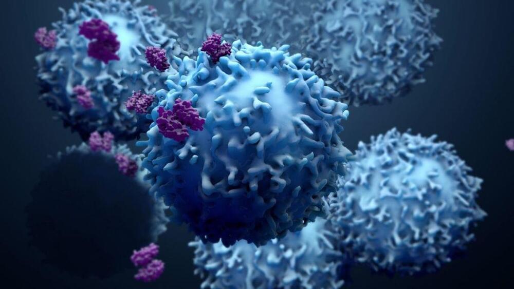
Myeloid cells are a population of cells classified to denote a specific lineage. “Myeloid” specifically refers to granulocytes and monocytes generated from the bone marrow. Many cells under this term share common progenitors from which they derive including, macrophages, neutrophils, basophils, and eosinophils. In the context of cancer, many of these cells become ‘pro-tumorigenic’. More specifically, they suppress the immune system to allow the tumor to proliferate and progress. Each myeloid cell type is associated with antitumor immune suppression. Myeloid cells suppress antitumor immune activity by blocking T cell activation, aid in angiogenesis (blood vessel formation) to increase metastasis, and producing cytokines or proteins that activate suppressive activity in other cells. Unfortunately, myeloid cells make up a major percentage within the tumor microenvironment, so targeting these cells is crucial. Many researchers are currently working on different ways to target these cell populations.

A recent article in Nature by Dr. Miriam Merad and her team demonstrated how protein signaling drives pro-tumor myeloid cell generation. Merad is a physician scientist, Director of the Precision Immunology Institute at Mount Sinai School of Medicine in New York, and Director of the Mount Sinai Human Immune Monitoring Center (HIMC). While her work focuses on targeting myeloid cells (particularly macrophages) to lower their suppressive phenotype and improve cancer treatment, her current publication identifies specific drivers of immunosuppressive myeloid states, previously undefined.

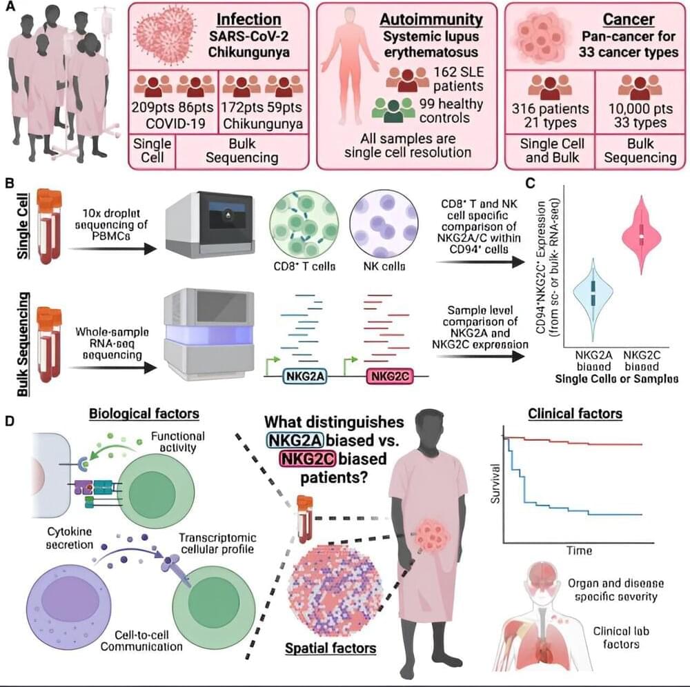
Merad and her team used advanced single cell sequencing to analyze non-small cell lung cancer (NSCLC) lesions from both humans and mice. Single cell sequencing is commonly used to identified up-and downregulated genes in a variety of cell types. By sequencing the tumor lesions the team discovered that interleukin 4 (IL-4) was a predictive driver of macrophages that infiltrated the tumor. Researchers used various genetically modified mouse models to conclude that the IL-4 receptor is necessary for tumor progression. Interestingly, they concluded that deletion of the IL-4 receptor in the progenitor phase reduced tumor growth compared to IL-4 deletion in mature macrophages which had little effect.

Cervical cancer, which develops in the lower portion of the uterus, known as the cervix, typically grows slowly. Cells in the cervix can change, a process known as dysplasia, and the resulting abnormal cells, if not removed, can develop into cervical cancer.

When cervical cancer occurs, treatment options vary based on many factors, including how far the disease has advanced and the overall health of the patient. Some cervical cancer patients may undergo a simple hysterectomy (also known as a complete or total hysterectomy). A simple hysterectomy removes the uterus and cervix.

The perimetrium, fat and connective tissue around the uterus, connects the uterus to the cervix and other tissues. Some patients with cervical cancer experience “parametrial invasion” (also referred to as parametrial infiltration), during which cervical cancer has spread into the parametrial tissue.