Graphene has long been regarded as one of the most promising materials for future electronics, but its relatively weak electron interactions have limited its potential for applications such as high-temperature superconductivity. Now, researchers from Tohoku University have overcome a major obstacle by creating a stable version of the long-sought “boron graphene” on the surface of a three-dimensional crystal, revealing a new quantum state that could lead to more energy-efficient electronic devices. The findings were published in Science Advances on July 2, 2026.
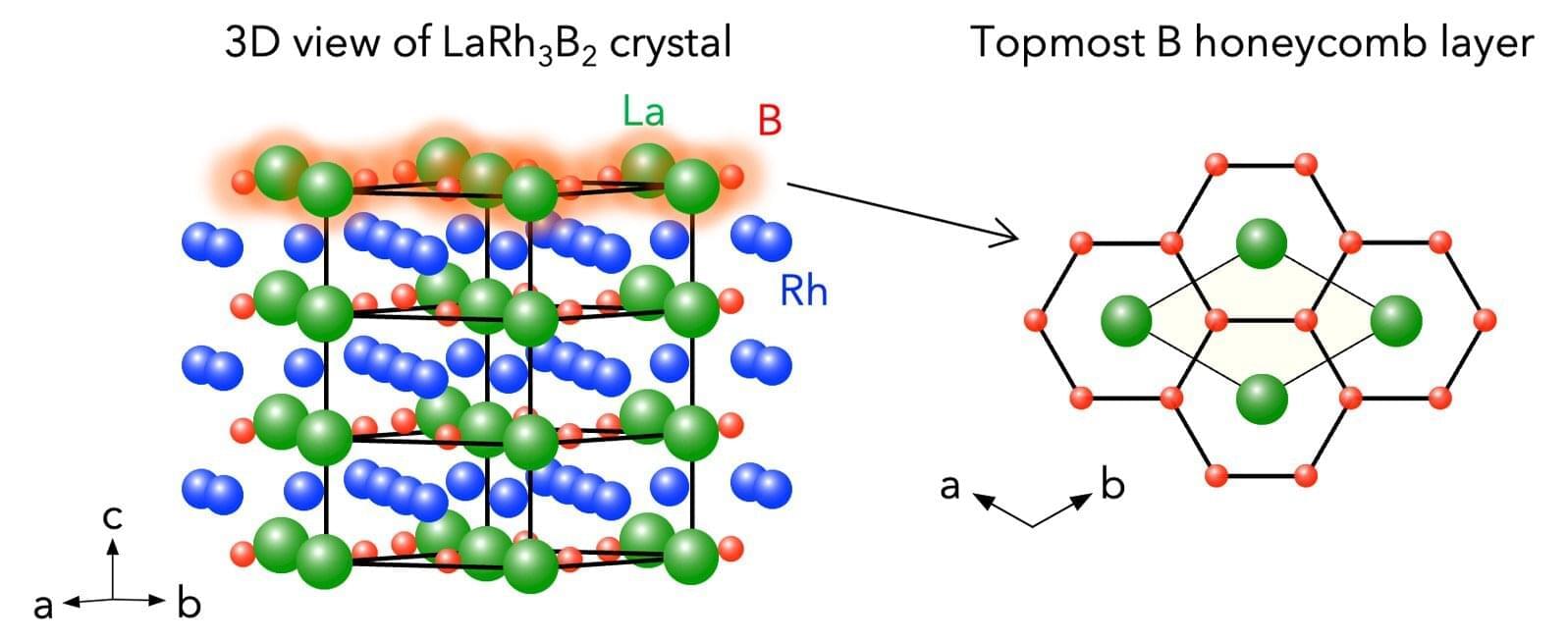
“We demonstrated a fundamentally new way of creating two-dimensional quantum materials,” says Takafumi Sato of Tohoku University’s Advanced Institute for Materials Research (WPI-AIMR). “Rather than attempting to produce an unstable free-standing sheet of boron atoms, we exposed a naturally occurring honeycomb boron layer that already exists within a stable three-dimensional crystal called LaRh3B2.”
For years, scientists have been interested in borophene—a two-dimensional sheet of boron atoms—because its stronger electron interactions could produce exotic quantum phenomena not seen in graphene. However, borophene’s ideal honeycomb structure is extremely unstable, making it almost impossible to manufacture.