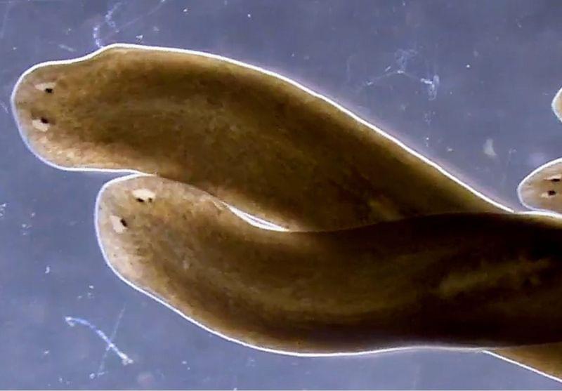
Stem cells in regenerating planarians don’t need their closest neighbors, overturning researchers’ understanding of the worms’ regenerative superpowers.
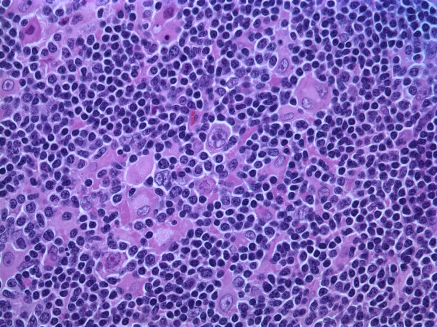
Cancer cells are known to reawaken embryonic genes to grow. A new study reveals the disease also hijacks the proteins, or “editors,” that control how those genes are read.
The findings, published in the journal Nucleic Acids Research, help explain why tumors grow so fast and adapt so well, and may point the way to new treatments.
Embryonic cells have to grow fast and must be able to transform into many different tissue types. The cells rely on genetic programs that are eventually switched off as tissues mature. Cancer reawakens these programs, giving the disease embryonic-like potential to fuel growth.

Oral cancer remains a serious health concern, often diagnosed too late for effective treatment, even though the mouth is easily accessible for routine examination. Dentists and dental hygienists are frequently the first to spot suspicious lesions, but many lack the specialized training to distinguish between benign and potentially malignant conditions.
To address this gap, researchers led by Rebecca Richards-Kortum at Rice University have developed and tested a low-cost, smartphone-based imaging system called mDOC (mobile Detection of Oral Cancer). Their recent study, published in Biophotonics Discovery, evaluates how well this system can help dental professionals decide when to refer patients to oral cancer specialists.
The mDOC device combines white light and autofluorescence imaging with machine learning to assess oral lesions. Autofluorescence imaging uses blue light to detect changes in tissue fluorescence, which can signal abnormal growth. However, this method alone can be misleading, as benign conditions like inflammation also reduce fluorescence.

Modern toads (Bufonidae) are among the most successful amphibians on the planet, a diverse group of more than 600 species that are found on every continent except Antarctica. But just how did they conquer the world? An international team of researchers set out to find the answer and discovered the toads’ global success was due to their toxic glands and geological timing.
Modern toads are a type of frog with a stout, squat body, relatively short legs, toothless mouths and a thick, dry, warty skin. One of their most distinctive features is a large gland behind each eye that secretes a poison to deter predators. They originated in South America and are found in diverse habitats like deserts and rainforests.
To find out how they got from South America to almost every other continent, the scientists analyzed fresh DNA samples from 124 species from Africa, Asia, Europe, South America, North America and Oceania. They combined this with existing genetic data from hundreds of other species. Using powerful computer models to process the genetic information, they traced the geological spread of toads over millions of years, identifying when survival features like their poisonous glands evolved and when they branched out to form new species.
Could tiny magnetic objects, that rapidly clump together and instantly fall apart again, one day perform delicate procedures inside the human body? A new study from researchers at the Max Planck Institute for Intelligent Systems in Stuttgart and at ETH Zurich introduces a wireless method to stiffen and relax small structures using magnetic fields, without wires, pumps, or physical contact.
In music, “jamming” refers to the spontaneous gathering of musicians who often improvise without aiming for a predefined outcome. In physics, jamming describes the transition of a material from a fluid-like to a solid-like state—like a traffic jam, where the flow of cars suddenly stops. This transformation can also be triggered on demand, offering a powerful and versatile way to control stiffness for robotic systems.
In most robotic applications, jamming is achieved using vacuum systems that suck air out of flexible enclosures filled with materials such as particles, fibers, or grains. But these systems require pumps, valves, and tubing—making them difficult to miniaturize.

A team from the Faculty of Physics and the Center for Quantum Optical Technologies at the University of Warsaw has developed a new type of all-optical radio receiver based on the fundamental properties of Rydberg atoms. The new type of receiver is not only extremely sensitive, but also provides internal calibration, and the antenna itself is powered only by laser light.
The results of the work, in which Sebastian Borówka, Mateusz Mazelanik, Wojciech Wasilewski and Michał Parniak participated, were published in Nature Communications. They open a new chapter in the technological implementation of quantum sensors.
In today’s society, huge amounts of digital information are transmitted around us every second. Much of it is transmitted by radio, i.e. using electromagnetic waves. For a very long time, amplitude modulation has been used to encode information, sending stronger and weaker waves.

A new twist on a classic material could advance quantum computing and make modern data centers more energy efficient, according to a team led by researchers at Penn State.
Barium titanate, first discovered in 1941, is known for its powerful electro-optic properties in bulk, or three-dimensional, crystals. Electro-optic materials like barium titanate act as bridges between electricity and light, converting signals carried by electrons into signals carried by photons, or particles of light.
However, despite its promise, barium titanate never became the industry standard for electro-optic devices, such as modulators, switches and sensors. Instead, lithium niobate—which is more stable and easier to fabricate, even if its properties don’t quite measure up with those of barium titanate—filled that role instead. But by reshaping barium titanate into ultrathin strained thin films, this could change, according to Venkat Gopalan, Penn State professor of materials science and engineering and co-author of the study published in Advanced Materials.
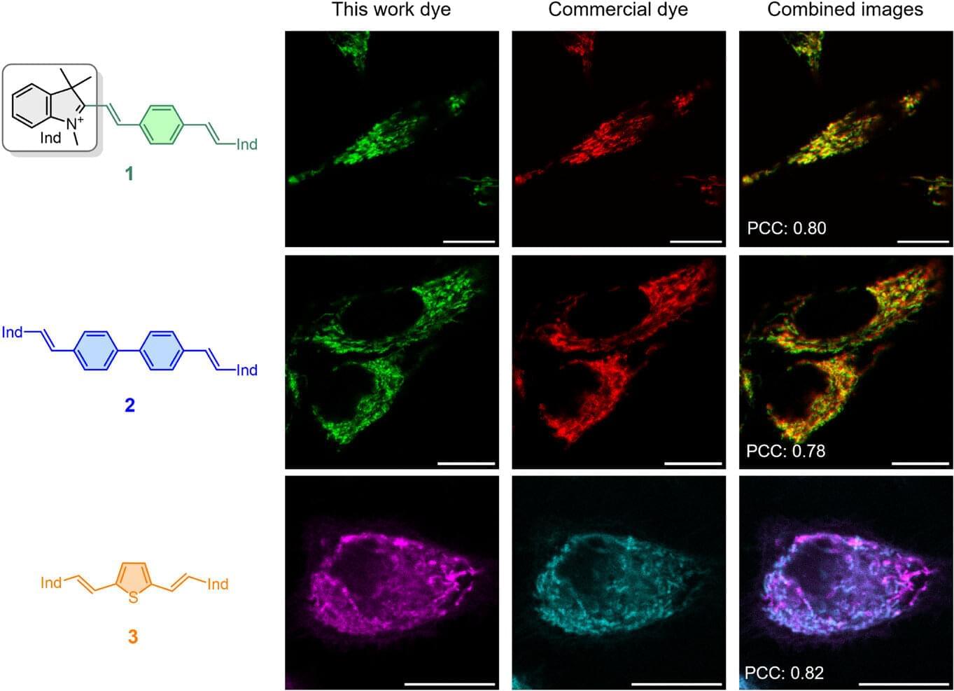
A team of researchers at the Departments of Physical Chemistry and Organic Chemistry of the University of Malaga and The Biomimetic Dendrimers and Photonic Laboratory of the research institute IBIMA Plataforma BIONAND has achieved a breakthrough that combines materials science and biomedicine. They have developed a new family of fluorescent molecules with promising applications in the study of living cells and the medicine of the future. The study has just been published in Advanced Materials.
The team of researchers has created a new family of fluorescent molecules that glow in a surprising way. These types of molecules typically lose part of their intensity or change to more dull colors when dissolved in water or other biological media. However, these new molecules do just the opposite: They emit a higher fluorescence intensity because their coloration shifts to the blue region of the light spectrum.
This behavior, which scientists described as “counterintuitive,” is key because it means that dyes work better in aqueous media like the inside of a cell, something essential for biomedical applications. In other words, they do not turn off when they are needed most but rather maintain—and even enhance—their brightness in real conditions of use.
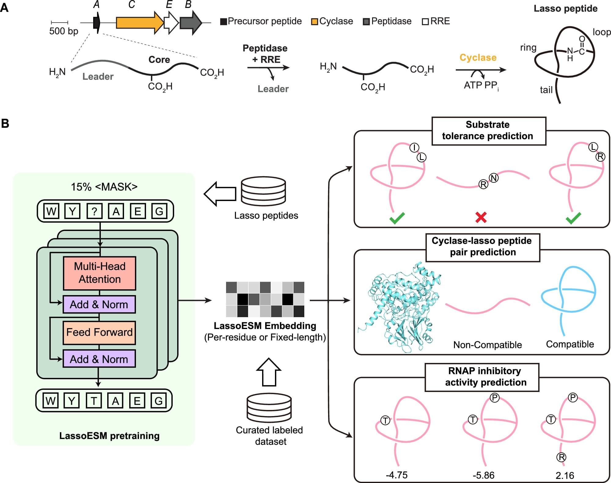
In the hunt for new therapeutics for cancer and infectious diseases, lasso peptides prove to be a catch. Their knot-like structures afford these molecules high stability and diverse biological activities, making them a promising avenue for new therapeutics. To better unleash their clinical potential, a team from the Carl R. Woese Institute for Genomic Biology has developed LassoESM, a new large language model for predicting lasso peptide properties.
The collaborative study was recently published in Nature Communications.
Lasso peptides are natural products made by bacteria. To produce these peptides, bacteria use ribosomes to build chains of amino acids that are then folded by biosynthetic enzymes into a unique slip knot-like structure. Through this process, thousands of different lasso peptides are generated, many of which have demonstrated antibacterial, antiviral, and anticancer properties.
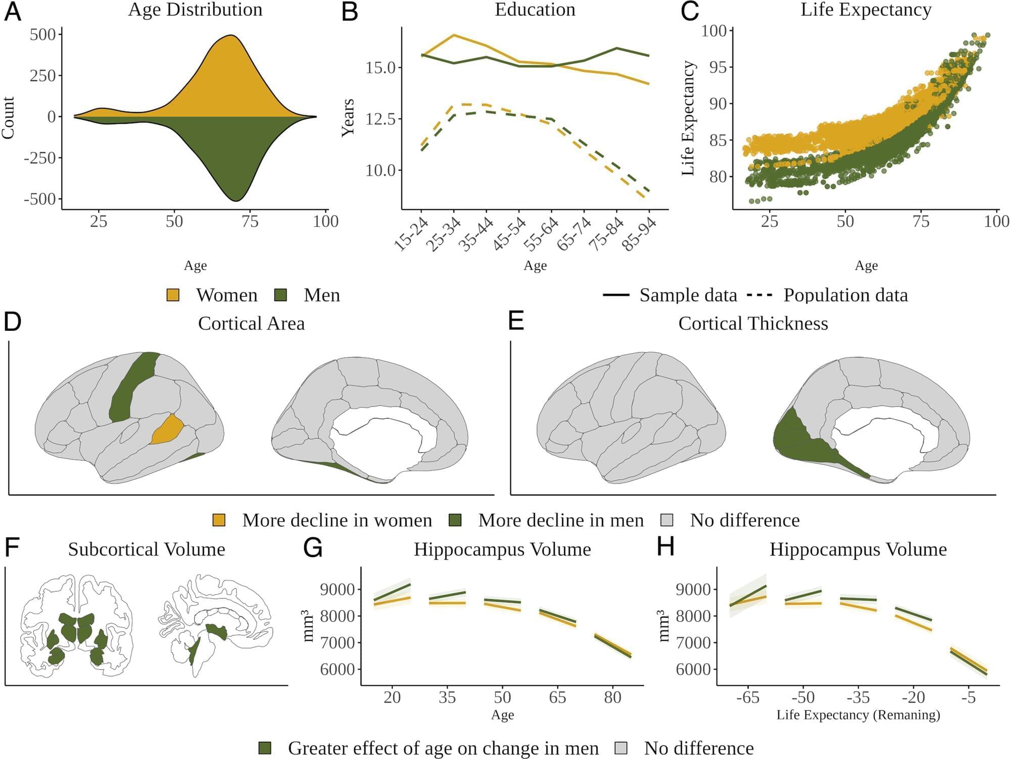
Women are far more likely than men to end up with Alzheimer’s disease (AD). This may, at least partially, be due to women’s longer average lifespans, but many scientists think there is probably more to the story. It would be easy to surmise that the increased risk is also related to differences in the way men’s and women’s brains change as they age. However, the research thus far has been unclear, as results across different brain regions and methods have been inconsistent.
Now, a new study, published in Proceedings of the National Academy of Sciences, indicates that it’s men who experience greater decline in more regions of the brain as they age. Researchers involved in the study analyzed 12,638 brain MRIs from 4,726 cognitively healthy participants (at least two scans per person) from the ages of 17–95 to find how age-related changes occurred and whether they differed between men and women.
The results showed that men experienced declines in cortical thickness and surface area in many regions of the brain and a decline in subcortical structures in older age. Meanwhile, women showed greater decline only in a few regions and more ventricular expansion in older adults. So, while differences in brain aging between the sexes are apparent, the cause of increased AD prevalence in women is still a bit mysterious.