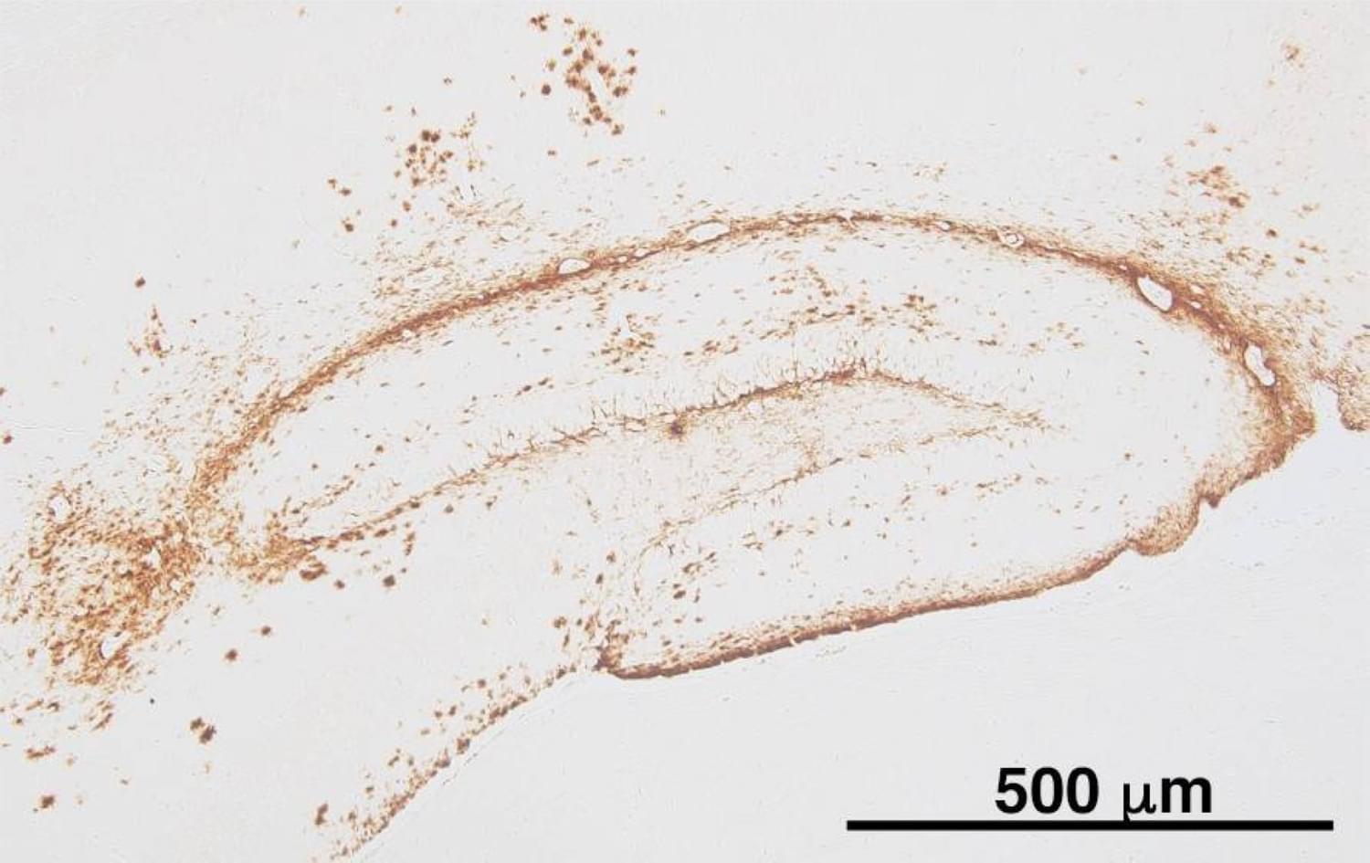
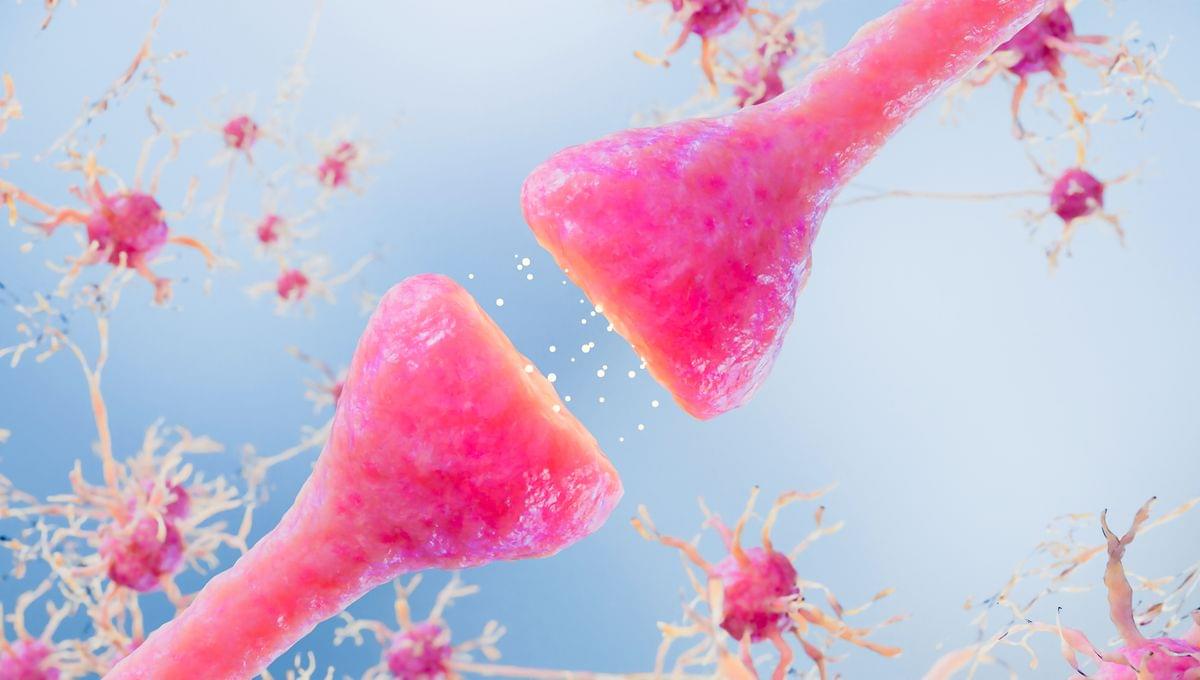
The discovery of a new seed of the amyloid beta protein could lead to early-stage therapy for neurodegenerative disease, according to researchers.

The MHRA’s latest alert warned about the dangers of unlicensed and counterfeit botulinum toxin products. “We also strongly urge the public to avoid unlicensed products and seek treatment only from appropriately qualified practitioners,” Cave said.
The agency said that products obtained outside regulated supply chains “do not meet UK safety and quality standards and can significantly increase the risk of serious side effects”. The agency also said that anyone considering treatment should ensure their practitioner is appropriately qualified, and confirm that the product being used is authorised for UK use.
The MHRA said it had worked with manufacturers to update product information, including patient leaflets, so that the risk of iatrogenic botulism is more clearly highlighted. It urged the public to report suspected side effects through the MHRA’s Yellow Card scheme.

No matter the company, AI chatbots were raving about the same guy named Elias Thorne. He must be pretty fascinating. And he is, at least on paper. Depending on the AI, he’s a lighthouse keeper, a clockmaker, a librarian, an explorer, and the star of countless stories. He’s appeared in books, music listings, YouTube videos, and even health guides. You’d think he was one of the most influential men on the planet.
But he doesn’t exist.
According to reporting by fine folks at 404 Media, researchers at Cornell University may have figured out why large language models invent and keep telling tales of the same fictional man. In a study examining roughly 20,000 AI-generated stories from all the big LLM models, including OpenAI, Anthropic, and Google, the research team found that the same handful of names and occupations kept cropping up. Specifically, names and words like Elias, Mara, Elara, lighthouse keeper, clockmaker, and librarian showed up in 88 percent of stories. Elias the lighthouse keeper appeared in nearly two-thirds of them.

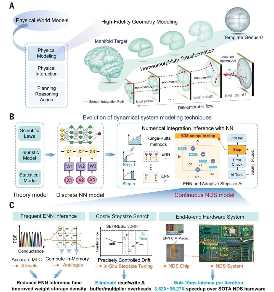
A research team has developed the world’s first chip that can match the speed at which the human brain functions. The study, titled “A sub–10-millisecond neural dynamical system based on phase-change memristors,” was published in Science and was led by Professor Yang Yuchao of Peking University, together with researchers from the Shanghai Institute of Microsystem and Information Technology, Chinese Academy of Sciences.
Neural dynamical systems combine neural networks with mathematical equations that describe how complex systems change over time. They are useful for physical modeling, medical imaging and three-dimensional brain reconstruction. However, these systems require repeated calculations, error checks and adjustments to the size of each calculation step. In conventional computers, data must also move frequently between memory and the processor, increasing processing time and energy use.
Fast and accurate brain modeling is important for technologies that must respond in real time, including brain–computer interfaces, surgical navigation and medical imaging. Existing hardware often requires too much time and power for these demanding calculations. By performing key operations directly in memory, the new chip reduces data movement and brings high-quality brain modeling closer to real-time use.
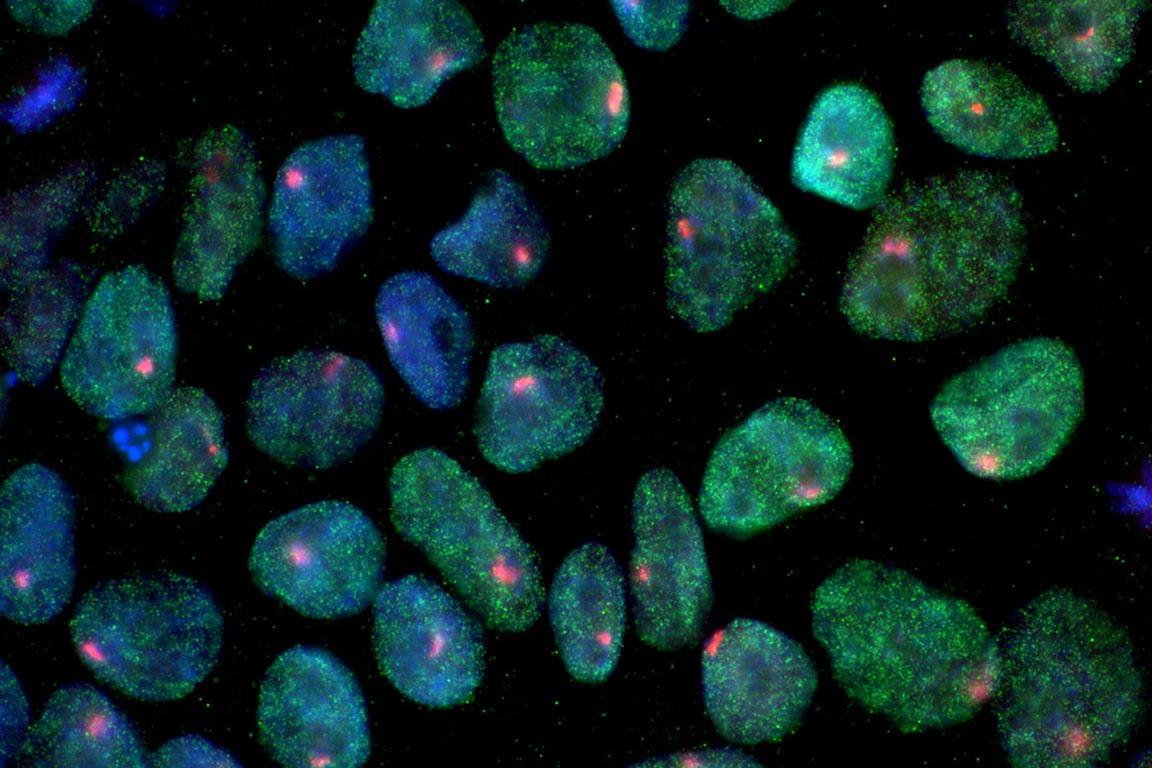
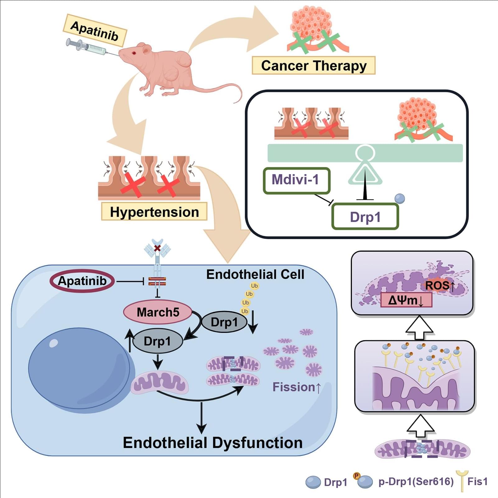
BACKGROUND: Apatinib is a tyrosine kinase inhibitor used for targeted cancer therapy, but its cardiovascular toxicity, particularly hypertension, limits its clinical application. We observed significant mitochondrial fragmentation in endothelial cells after apatinib treatment. This study aims to investigate the role of endothelial mitochondrial fission mediated by Drp1 (dynamin-related protein 1) in apatinib-induced hypertension. METHODS: We established an apatinib-targeted gastric cancer–bearing nude mice model. Apatinib was also administered to human umbilical vein endothelial cells in vitro. Mitochondrial morphology changes in endothelial cells were examined. The role of Drp1 in this process was validated using various experimental methods.