Toggle light / dark theme

Consciousness is comprised of arousal (i.e., wakefulness) and awareness. Substantial progress has been made in mapping the cortical networks that modulate awareness in the human brain, but knowledge about the subcortical networks that sustain arousal is lacking. We integrated data from ex vivo diffusion MRI, immunohistochemistry, and in vivo 7 Tesla functional MRI to map the connectivity of a subcortical arousal network that we postulate sustains wakefulness in the resting, conscious human brain, analogous to the cortical default mode network (DMN) that is believed to sustain self-awareness. We identified nodes of the proposed default ascending arousal network (dAAN) in the brainstem, hypothalamus, thalamus, and basal forebrain by correlating ex vivo diffusion MRI with immunohistochemistry in three human brain specimens from neurologically normal individuals scanned at 600–750 µm resolution. We performed deterministic and probabilistic tractography analyses of the diffusion MRI data to map dAAN intra-network connections and dAAN-DMN internetwork connections. Using a newly developed network-based autopsy of the human brain that integrates ex vivo MRI and histopathology, we identified projection, association, and commissural pathways linking dAAN nodes with one another and with cortical DMN nodes, providing a structural architecture for the integration of arousal and awareness in human consciousness. We release the ex vivo diffusion MRI data, corresponding immunohistochemistry data, network-based autopsy methods, and a new brainstem dAAN atlas to support efforts to map the connectivity of human consciousness.

One sentence summary We performed ex vivo diffusion MRI, immunohistochemistry, and in vivo 7 Tesla functional MRI to map brainstem connections that sustain wakefulness in human consciousness.

BF has a financial interest in CorticoMetrics, a company whose medical pursuits focus on brain imaging and measurement technologies. BF’s interests were reviewed and are managed by Massachusetts General Hospital and Mass General Brigham HealthCare in accordance with their conflict-of-interest policies.
Is consciousness a scientific problem to be solved? Or a philosophical problem that will remain a mystery? What do scientists who study the brain think? And why do they think the way they do? These leading brain scientists share their intimate ideas about how the brain generates consciousness.

Free access to Closer to Truth’s library of 5,000 videos: http://bit.ly/376lkKN

Watch more interviews on neuroscience and the hard problem of consciousness: https://bit.ly/3NZ2gn5

Arnold B. Scheibel was a Professor of Neurobiology and Psychiatry and former Director of the Brain Research Institute (BRI) at UCLA.

Learn more about the Cognitive Science Student Association and the California Cognitive Science Conference at https://cssa.berkeley.edu.

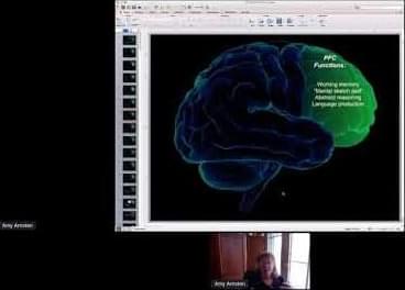
Amy Arnsten — Yale University.

Abstract.
The recently evolved prefrontal cortex (PFC) subserves many of our highest-order cognitive functions, generating and sustaining the mental representations that underlie working memory, abstract reasoning, and top-down control of thought, action, and emotion. Due to the pioneering research of Patricia Goldman-Rakic, we have learned much about the neural basis underlying the ability of the dorsolateral prefrontal cortex (dlPFC) to generate mental representations, where microcircuits in deep layer III have extensive recurrent excitatory connections to maintain neuronal firing in the absence of sensory stimulation, while GABAergic interneurons provide lateral inhibition to refine the contents of working memory. However, these dlPFC circuits are also tremendously dependent on arousal state, with a narrow inverted U response to levels of acetylcholine, dopamine and norepinephrine. Even quite mild uncontrollable stress increases the release of dopamine and norepinephrine in the PFC, which rapidly impairs PFC functioning by 1) stimulating D1 and alpha-1-receptors, respectively, 2) these, in turn, activate feedforward calcium-cAMP signaling within spines, which then 3) open nearby potassium channels to disconnect PFC networks and take the PFC “off-line”. With chronic stress exposure, there is actual atrophy of PFC dendrites and spines. Understanding the neural events that weaken vs. strengthen PFC connectivity and function has led to the development of treatments for patients with stress-related PFC dysfunction, e.g. guanfacine and prazosin. This knowledge is also helping to illuminate the etiology of cognitive disorders, as genetic insults in schizophrenia often increase the activity of these stress signaling pathways, and the molecules that regulate the stress signaling pathways are lost with advancing age, leading to tau pathology as seen in Alzheimer’s disease.

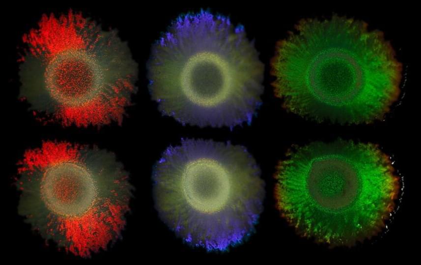
An international team of researchers of the Cluster of Excellence “Balance of the Microverse” at the University of Jena has investigated the mechanism that makes some types of bacteria reflect light without using pigments. The researchers were interested in the genes responsible and discovered important ecological connections. Their findings appear in the Proceedings of the National Academy of Sciences.

The iridescent colors known from peacock feathers or butterfly wings are created by tiny structures that reflect light in a special way. Some form similar glittering structures.

In collaboration with the Max Planck Institute of Colloids and Interfaces, Leibniz Institute DSMZ-German Collection of Microorganisms and Cell Cultures, Utrecht University, University of Cambridge, and the Netherlands Institute for Sea Research, the scientists sequenced the DNA of 87 structurally colored bacteria and 30 colorless strains and identified genes that are responsible for these fascinating colonies. These findings could lead to the development of environmentally-friendly dyes and materials, a key interest of the collaborating biotechnology company Hoekmine BV.

Scientists have developed a new way to trap small particles with light. Building on the Nobel Prize winning technique of optical tweezers (Arthur Ashkin, 2018), a team of physicists, led by Dr. David Phillips at the University of Exeter, has advanced the possibilities of optical trapping.

The research paper, published in the journal Science Advances, is titled “Photon-efficient optical via wavefront shaping.”

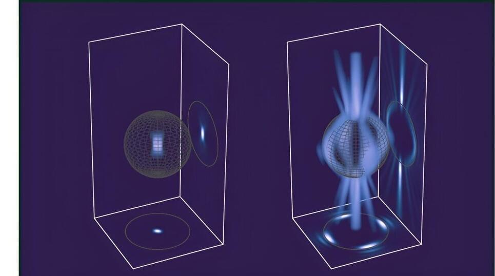
Conventional optical tweezers, developed in the 1980s, are a tightly focused laser beam which can attract and trap certain micro-sized particles or organisms, akin to grabbing something with a pair of tweezers.

A levitating microparticle is observed to recoil when a nucleus embedded in the particle decays—opening the door to future searches of invisible decay products.

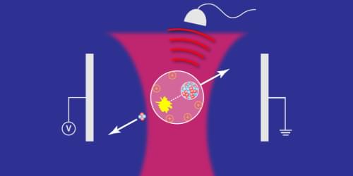
For centuries, physicists have exploited momentum conservation as a powerful means to analyze dynamical processes, from billiard-ball collisions to galaxy formation to subatomic particle creation in accelerators. David Moore and his research team at Yale University have now put this approach to work in a new setting: they used momentum conservation to determine when a radioactive atom emitted a single helium nucleus, known as an alpha particle (Fig. 1) [1]. The demonstration suggests that—with further improvements—researchers might be able to use this technique to detect other nuclear-decay products, such as neutrinos and hypothetical dark-matter particles (see also Special Feature: Sensing a Nuclear Kick on a Speck of Dust).

The basic idea is simple: if the radioactive atom is embedded in a larger object, then an outgoing decay product will exert a backreaction on that object, causing it to recoil in the opposite direction. But is it really possible to detect the recoil kick from a particle as small as a helium nucleus? The answer lies in how precisely we can measure the larger object’s momentum. One of the main limitations is friction: if the larger object is slowed down by frictional forces, then its motion won’t reflect the impulse from the decaying particle.