Acetic acid, also known as acetate, and other products that can be developed from acetic acid are used in a variety of industries, from food production to medicine to agriculture. Currently, acetate production uses a significant amount of energy and results in harmful waste products. The efficient and sustainable production of acetate is an important target for researchers interested in improving industrial sustainability.
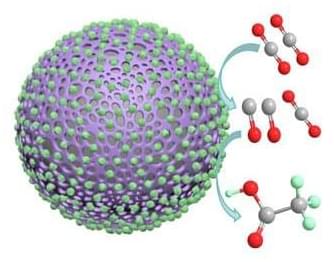
A paper published in Carbon Future (“CO 2 electroreduction to acetate by enhanced tandem effects of surface intermediate over Co 3 O 4 supported polyaniline catalyst”) outlines a method using a polyaniline catalyst with cobalt oxide nanoparticles to produce acetate through carbon dioxide electroreduction.
This image shows a polyaniline catalyst coated in cobalt oxide nanoparticles and demonstrates how the catalyst facilitates the conversion of carbon dioxide to carbon monoxide to acetate. (Image: Carbon Future)
