Toggle light / dark theme

Newly Discovered Features Of Collagen May Help Shed Light On Disease Processes

Interesting.


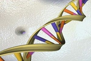
WHAT: Scientists at the National Institutes of Health are reporting new, unexpected details about the fundamental structure of collagen, the most abundant protein in the human body. In lab experiments, they demonstrated that collagen, once viewed as inert, forms structures that regulate how certain enzymes break down and remodel body tissue. The finding of this regulatory system provides a molecular view of the potential role of physical forces at work in heart disease, cancer, arthritis, and other disease-related processes, they say. The study appears in the current online issue of the Proceedings of the National Academy of Sciences.

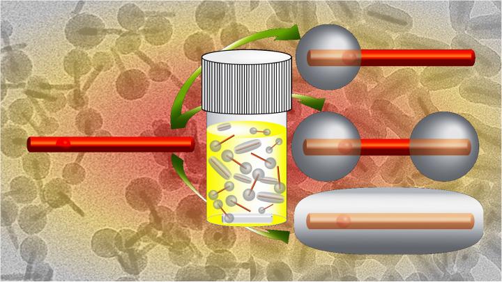
Scientists have known for years that collagen remodeling plays an important role in a wide variety of biological processes ranging from wound healing to cancer growth. In particular, researchers know that collagen is broken down by a certain class of enzymes called matrix metalloproteinases (MMPs), but exactly how they did this remained somewhat of a mystery, until now.

In the NIH study, the scientists isolated individual, nano-sized collagen fibrils from rat-tail tendons. They then exposed the collagen fibrils to fluorescently-labeled human MMP enzymes. Using video microscopy, the scientists tracked thousands of enzymes moving along a fibril. Unexpectedly, the scientists observed that the enzymes preferred to attach at certain sites along the fibril, and over time these attachment sites slowly moved, or disappeared and reappeared in other positions. These observations revealed collagen fibrils have defects that spontaneously form and heal. In the presence of tension, such as when tendons stretch, defects are likely eliminated, preventing enzymes from breaking down collagen that is loaded by physical force, the researchers suggest. In short, they identified a possible strain-sensitive mechanism for regulating tissue remodeling.

Germs add ripples to make ‘groovy’ graphene

Graphene, a two-dimensional wonder-material composed of a single layer of carbon atoms linked in a hexagonal chicken-wire pattern, has attracted intense interest for its phenomenal ability to conduct electricity. Now University of Illinois at Chicago researchers have used rod-shaped bacteria — precisely aligned in an electric field, then vacuum-shrunk under a graphene sheet — to introduce nanoscale ripples in the material, causing it to conduct electrons differently in perpendicular directions.

The resulting material, sort of a graphene nano-corduroy, can be applied to a silicon chip and may add to graphene’s almost limitless potential in electronics and nanotechnology. The finding is reported in the journal ACS Nano.

“The current across the graphene wrinkles is less than the current along them,” says Vikas Berry, associate professor and interim head of chemical engineering at UIC, who led the research.

Atomic bits despite zero-point energy? Jülich scientists explore novel ways of developing stable nanomagnets

Stable nanomagnets that ultimately improves data storage on the smallest of devices.


Abstract: So-called “zero-point energy” is a term familiar to some cinema lovers or series fans; in the fictional world of animated films such as “The Incredibles” or the TV series “Stargate Atlantis”, it denotes a powerful and virtually inexhaustible energy source. Whether it could ever be used as such is arguable. Scientists at Jülich have now found out that it plays an important role in the stability of nanomagnets. These are of great technical interest for the magnetic storage of data, but so far have never been sufficiently stable. Researchers are now pointing the way to making it possible to produce nanomagnets with low zero-point energy and thus a higher degree of stability (Nano Letters, DOI: 10.1021/acs.nanolett.6b01344).

Since the 1970s, the number of components in computer chips has doubled every one to two years, their size diminishing. This development has made the production of small, powerful computers such as smart phones possible for the first time. In the meantime, many components are only about as big as a virus and the miniaturization process has slowed down. This is because below approximately a nanometre, a billionth of a meter in size, quantum effects come into play. They make it harder, for example, to stabilise magnetic moments. Researchers worldwide are looking for suitable materials for magnetically stable nanomagnets so that data can be stored safely in the smallest of spaces.

In this context, stable means that the magnetic moments point consistently in one of two preassigned directions. The direction then codes the bit. However, the magnetic moments of atoms are always in motion. The trigger here is the so-called zero-point energy, the energy that a quantum mechanical system possesses in its ground state at absolute zero temperature. “It makes the magnetic moments of atoms fluctuate even at the lowest of temperatures and thus works against the stability of the magnetic moments”, explains Dr. Julen Ibañez-Azpiroz, from the Helmholtz Young Investigators Group “Functional Nanoscale Structure Probe and Simulation Laboratory” at the Peter Grünberg Institute and at the Institute for Advanced Simulation. When too much energy exists within the system, the magnetic moments turn over and the saved information is lost.

‘Nano scalpel’ allows structuring of samples with nanometre precision

A new “nano scalpel” enables scientists at DESY to prepare samples or materials with nanometre precision while following the process with a scanning electron microscope. The Focused Ion Beam, or FIB, microscope which has now gone into service also allows a detailed view of the inner structure of materials. The device was purchased by the University of Bayreuth, as part of a joint research project on the DESY campus funded by the Federal Ministry of Research. The FIB will be operated at the DESY NanoLab jointly with the University of Bayreuth.

“The microscope is not only able to examine microscopic defects, cracks or point-like corrosion sites underneath the surfaces of , but also to machine the surface of samples with extremely high precision, on a nanometre scale,” explains Maxim Bykov, project scientist from the University of Bayreuth. A nanometre is a millionth of a millimetre. The can be used to remove material as though it were a microscopic milling machine; as a result, the combined ion beam and electron microscope is particularly interesting for a wide range of applications in nanotechnology, materials science and biology.

“Apart from examining the structure of materials, the ability of the ion beam to remove material also leads to a wide range of different applications,” says Natalia Dubrovinskaia who is a professor at the University of Bayreuth and in charge of the joint research project (No. 05K13WC3). One example is the preparation of tiny diamond anvils, which are used to hold samples during ultra high-pressure experiments. The diamonds used for this are so small that there is no other way of preparing them. The ion beam allows so-called double-staged diamond anvil cells to be prepared with nanometre precision. The ultra high-pressure experiments are carried out at DESY’s Extreme Conditions Beamline (ECB) P02.2, headed by DESY scientist Hanns-Peter Liermann.

Researchers develop faster, precise silica coating process for quantum dot nanorods

Faster and better method around Q-dots development which ultimately extends the quality of Quantum Dots plus mass production of Q-Dots is much faster through this new method. Hoping this causes the costs of new cameras, phone displays, monitors/ video displays are now able to be created more cheaply and in larger quantities.


Materials researchers at North Carolina State University have fine-tuned a technique that enables them to apply precisely controlled silica coatings to quantum dot nanorods in a day — up to 21 times faster than previous methods. In addition to saving time, the advance means the quantum dots are less likely to degrade, preserving their advantageous optical properties.

Quantum dots are nanoscale semiconductor materials whose small size cause them to have electron energy levels that differ from larger-scale versions of the same material. By controlling the size of the quantum dots, researchers can control the relevant energy levels — and those energy levels give quantum dots novel optical properties. These characteristics make quantum dots promising for applications such as opto-electronics and display technologies.

But quantum dots are surrounded by ligands, which are organic molecules that are sensitive to heat. If the ligands are damaged, the optical properties of the quantum dots suffer.

A Flexible Evolving Approach To Computing

To truly reach a fully connected world/ singularity we have to move tech into more and more bio-computing world. I do believe QC will assist us in getting the fundamental infrastructure we need for singularity.


We already must deal with computers too much rather than too little, and there is already lots of advanced computing done also for example in materials science and nanotechnology, for example molecular dynamics (MD) and Monte Carlo simulations.[2] The molecular biologist’s programs for predicting protein folding can also count as nanotechnology. Nevertheless, all of our previous articles concluded that we need more computing, and several mentioned statistics. This would sound predictable if coming from a statistical physicist with a background in computing, advertising his skills. However, we mean a more efficient computing rather than simply more.

We started the type of computing we do only recently and for reasons not yet mentioned: Given complex nano-micro compounds, materials’ characterization is difficult due to the three-dimensional complexity of the structures. We originally integrated image analysis with simulation in order to derive 3D structure from 2D images (SEM) and projections (TEM).[3,4] The most fruitful result was however the insight into how easy it is to create adaptable software that analyzes images and keeps track of all the data, calculating anything desired such as comparisons with numerical simulations, all in one integrated system.[5,6] Many of the previously discussed issues, for example error reporting, are thereby basically already automatically solved!

Adapting software sounds prohibitively difficult: Who in my lab can modify software? Nowadays everybody! Today, programming is done partially graphically, for example with LabView™, where no programming language appears anymore. We work with Mathematica and therefore with programming code, but we mostly just download parts of code and adapt them playfully until they behave as desired. To whomever such does not count as the ability to program, we cannot program!