Toggle light / dark theme

Histology is used to identify structural details of tissue at the microscale in the pathology lab, but analyses remain two-dimensional (2D) as they are limited to the same plane. Nondestructive 3D technologies including X-ray micro and nano-computed tomography (nanoCT) have proven validity to understand anatomical structures, since they allow arbitrary viewing angles and 3D structural detail. However, low attenuation of soft tissue has hampered their application in the field of 3D virtual histology. In a recent study, now published on Scientific Reports, Mark Müller and colleagues at the Department of Physics and Bioengineering have developed a hematein-based X-ray staining method to specifically target cell nuclei, followed by demonstrations on a whole liver lobule of a mouse.


Scientists have created a rubbery, shape-shifting material that morphs from one sophisticated form to another on demand.

The shapes programmed into a polymer appear in ambient conditions and melt away when under heat. The process also works in reverse.

The smooth operation belies a battle at the nanoscale, where liquid crystals and the elastomer in which they’re embedded fight for control. When cool, the shape programmed into the liquid crystals dominates, but when heated, the crystals relax within the rubber band-like elastomer, like ice melting into water.

Read more

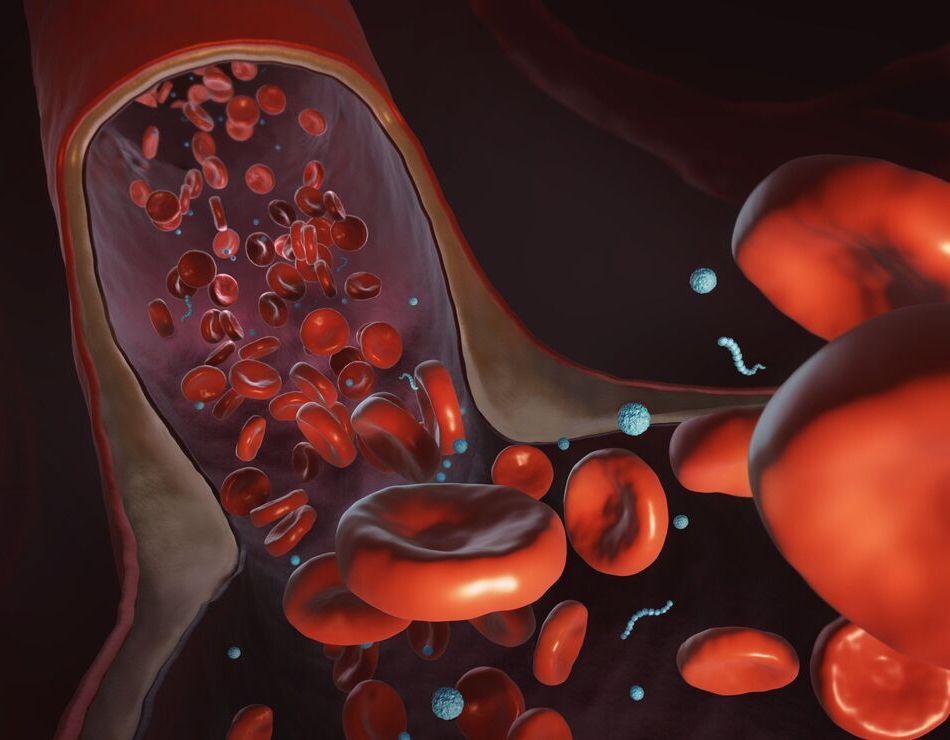
Coming up with potent anti-cancer drugs is one thing, delivering them to the site of a tumor inside the body is very much another. With a complicated organism guarded by a highly evolved immune system to navigate, getting these particles to there target in one piece is a challenging task, and one that scientists are continuing to tackle from all angles. A promising new approach developed at Virginia Tech leans on the penetrative properties of a salmonella infection, which they’ve found can be used as a vehicle to smuggle cancer-fighting nanoparticles into a tumor in a huge abundance.

Read more

MIT researchers invented a method of shrinking objects to the nanoscale.

The team can generate structures one-thousandth the volume of the original using a variety of materials, including metals, quantum dots, and DNA.

Existing techniques—like etching patterns onto a surface with light—work for 2D nanostructures, but not 3D. And while it’s possible to make 3D nanostructures, the process is slow, challenging, and restrictive.

Read more

Nano-sized particles already make bicycles and tennis rackets lighter and stronger, protect eyeglasses from scratches, and help direct chemotherapy drugs to cancer cells. But their usefulness depends on being able to precisely sculpt them into the right configurations—no easy task when they’re so tiny that thousands of them could fit into the thickness of a sheet of paper.

Read more