Researchers have developed nano-scale drills that kill bacteria by drilling holes into their membranes. They are powered by visible light.


A new technology is using particles of gold to make colors. With further work, the method developed at Aalto University could herald a new display technology.
The technique uses gold nanocylinders suspended in a gel. The gel only transmits certain colors when lit by polarized light, and the color depends on the orientation of the gold nanocylinders. In a clever twist, a collaboration led by Anton Kuzyk’s and Juho Pokki’s research groups used DNA molecules to control the orientation of gold nanocylinders in the gel.
“DNA isn’t just an information carrier—it can also be a building block. We designed the DNA molecules to have a certain melting temperature, so we could basically program the material,” says Aalto doctoral candidate Joonas Ryssy, the study’s lead author. When the gel heats past the melting temperature, the DNA molecules loosen their grip and the gold nanocylinders change orientation. When the temperature drops, they tighten up again, and the nanoparticles go back to their original position.


Nanosheets are finely structured two-dimensional materials and have great potential for innovation. They are fixed on top of each other in layered crystals, and must first be separated from each other so that they can be used, for example, to filter gas mixtures or for efficient gas barriers. A research team at the University of Bayreuth has now developed a gentle, environmentally-friendly process for this difficult process of delamination that can even be used on an industrial scale. This is the first time that a crystal from the technologically attractive group of zeolites has been made usable for a broad field of potential applications.
The delamination process developed in Bayreuth under the direction of Prof. Dr. Josef Breu is characterized by the fact that the structures of the nanosheets isolated from each other remain undamaged. It also has the advantage that it can be used at normal room temperature. The researchers present their results in detail in Science Advances.
The two-dimensional nanosheets, which lie on top of each other in layered crystals, are held together by electrostatic forces. In order for them to be used for technological applications, the electrostatic forces must be overcome, and the nanosheets detached from each other. A method particularly suitable for this is osmotic swelling, in which the nanosheets are forced apart by water and the molecules and ions dissolved in it. So far, however, it has only been possible to apply it to a few types of crystals, including some clay minerals, titanates, and niobates. For the group of zeolites, however, whose nanosheets are highly interesting for the production of functional membranes due to their silicate-containing fine structures, the mechanism of osmotic swelling has not yet been applicable.

It’s a hot topic, at least on social media: tiny plastic particles allegedly end up not only in oceans and lakes, but also in drinking water—and, yes, even in bottled mineral water. Eawag and the Zurich Water Works launched a joint project in 2019 to find out whether the tiniest of particles, measuring less than a thousandth of a millimeter across, actually find their way from lake water into drinking water pipes and therefore into homes, hospitals and restaurants.
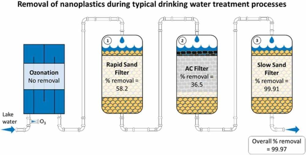
The results are now in, and they include some reassuring findings. In a report published today in the Journal of Hazardous Materials, the researchers show that even if untreated water contained considerable quantities of nanoplastics, these particles were retained in sand filters very efficiently during water treatment. Both in laboratory tests and in a larger test facility located directly on the premises of the Zurich Water Works, the biologically active slow sand filter was the most effective at retaining nanoparticles—achieving an efficacy level in the region of 99.9%.
So far, there has only been limited research into how exactly nanoplastics are formed. “But it would appear that the degradation of larger plastic particles in the environment eventually results in nanoplastics,” says Ralf Kägi, Head of Eawag’s Particle Laboratory. However, even the process of identifying nanoplastic particles is anything but easy. For this, the team of researchers from Eawag, ETH Zurich, EPFL and the Politecnico di Torino used labeled nanoplastic particles, whose route through—or final location in—the water treatment process could be tracked using a mass spectrometer. This process is similar to that used in medicine, where cancer cells are specifically labeled in order to monitor their potential distribution in the human body.
Once the tiny, iron-based particles are added to the water, the lithium is drawn out of the water and binds to them. Then with the help of a magnet, the nanoparticles can be collected in just minutes with the lithium hitching a ride, no longer suspended in the liquid and ready for easy extraction. After the lithium is extracted, the recharged nanoparticles can be used again.
New approach uses magnetic nanoparticles to extract valuable rare earth elements from geothermal fluids.

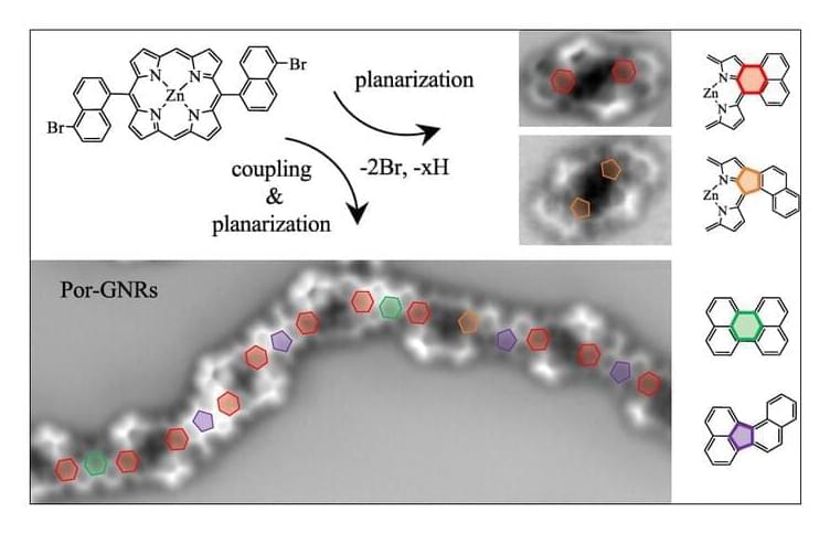
On-surface synthesis made the fabrication of extended, atomically precise π-conjugated nanostructures on solid supports possible, with graphene nanoribbons (GNRs) and porphyrin-derived oligomers standing out. To date, examples combining these two prominent material classes are scarce, even though the chemically versatile porphyrins and the atomistic details of the nanographene spacers promise an easy tunability of structural and functional properties of the resulting hybrid structures. Here, we report the on-surface synthesis of extended benzenoid-and nonbenzenoid-coupled porphyrin–graphene nanoribbon hybrids by sequential Ullmann-type and cyclodehydrogenation reactions of a tailored Zn(II) 5,15-bis(5-bromo-1-naphthyl)porphyrin (Por(BrNaph)2) precursor on Au(111) and Ag(111).


Circa 2020 Electricity free grow lights using quantum dot leds.
While costs are coming down for controlled environment agriculture, electricity remains one of the highest because it has to power the LEDs that provide the lighting formula for plant growth. But a materials science company called UbiQD wants to change that by replacing electricity with a more efficient means of lighting: quantum dots.
Quantum dots are semiconductor nanoparticles that can transport electrons. When exposed to UV lighting, these particles emit lights of various colors, and can be adjusted in size to emit a specific color. For example, larger particles emit redder wavelengths, while smaller ones shift to blue.
Via its UbiGro product, UbiQD uses a patented quantum dot technology to create a layer of lighting in greenhouses. Quantum dots are embedded into a film that is installed beneath a greenhouse cover. When illuminated by sunlight, the film converts shorter wavelengths (UV and blue) to longer ones (red/orange), the latter being the most photosynthetically efficient wavelengths.

From TVs, to solar cells, to cutting-edge cancer treatments, quantum dots are beginning to exhibit their unique potential in many fields, but manufacturing them at scale would raise some issues concerning the environment. Scientists at Japan’s Hiroshima University have demonstrated a greener path forward in this area, by using discarded rice husks to produce the world’s first silicon quantum dot LED light.
“Since typical quantum dots often involve toxic material, such as cadmium, lead, or other heavy metals, environmental concerns have been frequently deliberated when using nanomaterials,” said Ken-ichi Saitow, lead study author and a professor of chemistry at Hiroshima University. “Our proposed process and fabrication method for quantum dots minimizes these concerns.”
The type of quantum dots pursued by Saitow and his team are silicon quantum dots, which eschew heavy metals and offer some other benefits, too. Their stability and higher operating temperatures makes them one of the leading candidates for use in quantum computing, while their non-toxic nature also makes them suitable for use in medical applications.