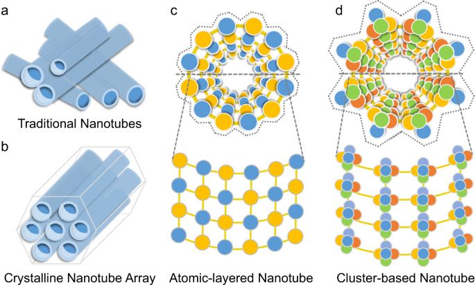
Interesting properties of carbon nanotubes prompt a search for diverse inorganic nanotubes. Here, the authors report a supertetrahedral chalcogenide cluster-based semiconducting nanotube array that exhibits high electric conductivity and oriented photoconductive behavior.
Check out our new promo for #transvision #future Summit 2021! Get your tickets! -> www.TransVisionMadrid.com There will be talks about #longevity #artificialintelligence #cryonics and much much more. You will also be able to network with speakers and attendees during 5 optional dinner/cocktails, and 2 tours of several UNESCO heritage sites and historical places: Ávila, Segovia, Monsaterio de El Escorial, Valle de los Caídos (Valley of the Fallen), Aranjuez & Toledo.
Humanity Plus Humanity Plus Humanity Plus Magazine MUTISHAN Interactive Vivian Francos #SEOHashtag Alcor Life Extension Foundation Cryonics Institute Cryonics Institute SENS Research Foundation SENS Research Foundation Posthuman Network Posthuman Network Cryonics4U Longevity Conferences Longevity for All U.S. Transhumanist Party Transhumanist Party Australia Transhumanist Party Virtual Rational Transhumanism Singularity University Ray Kurzweil Singularity Singularity Hub Ray Kurzweil’s Singularity Singularity Network Transhumanismo Brasil Transhumanismo Brasil TRANSHUMANISMO Christian Transhumanist Association Mormon Transhumanist Association SingularityNET Singularitarianism Foresight Institute Lifeboat Foundation Lifeboat Foundation Machine Intelligence Research Institute KrioRus The Hedonistic Imperative — Paradise Engineering.
Promo by sergio tarrero for alianza futurista & transvision madrid.
Spain will host the next world futurist summit on October 8, 9 and 10, 2021. Humanity+ will be the main international organizer of this international congress. Afterwards, during October 11 and 12, we will continue with informal conversations while traveling to UNESCO World Heritage Sites around Madrid: Aranjuez, Ávila, El Escorial, Segovia y Toledo. Every night will finish with optional cocktails in beautiful places for networking and meeting the participants and speakers.
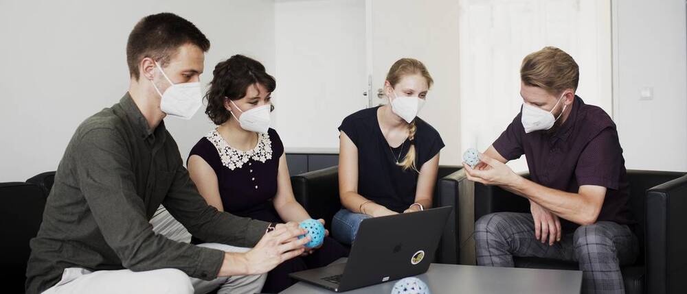
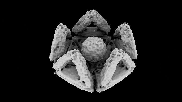
To date, there are no effective antidotes against most virus infections. An interdisciplinary research team at the Technical University of Munich (TUM) has now developed a new approach: they engulf and neutralize viruses with nano-capsules tailored from genetic material using the DNA origami method. The strategy has already been tested against hepatitis and adeno-associated viruses in cell cultures. It may also prove successful against corona viruses.
To date, there are no effective antidotes against most virus infections. An interdisciplinary research team at the Technical University of Munich (TUM) has now developed a new approach: they engulf and neutralize viruses with nano-capsules tailored from genetic material using the DNA origami method. The strategy has already been tested against hepatitis and adeno-associated viruses in cell cultures. It may also prove successful against coronaviruses.
There are antibiotics against dangerous bacteria, but few antidotes to treat acute viral infections. Some infections can be prevented by vaccination but developing new vaccines is a long and laborious process.
Now an interdisciplinary research team from the Technical University of Munich, the Helmholtz Zentrum München, and the Brandeis University (USA) is proposing a novel strategy for the treatment of acute viral infections: The team has developed nanostructures made of DNA, the substance that makes up our genetic material, that can trap viruses and render them harmless.
Circa 1999 could lead to a sorta room temperature hydrogen fill up.
Masses of single-walled carbon nanotubes (SWNTs) with a large mean diameter of about 1.85 nanometers, synthesized by a semicontinuous hydrogen arc discharge method, were employed for hydrogen adsorption experiments in their as-prepared and pretreated states. A hydrogen storage capacity of 4.2 weight percent, or a hydrogen to carbon atom ratio of 0.52, was achieved reproducibly at room temperature under a modestly high pressure (about 10 megapascal) for a SWNT sample of about 500 milligram weight that was soaked in hydrochloric acid and then heat-treated in vacuum. Moreover, 78.3 percent of the adsorbed hydrogen (3.3 weight percent) could be released under ambient pressure at room temperature, while the release of the residual stored hydrogen (0.9 weight percent) required some heating of the sample.
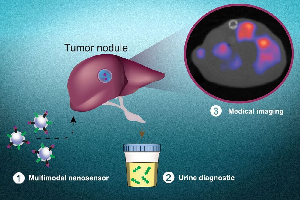
Most of the tests that doctors use to diagnose cancer — such as mammography, colonoscopy, and CT scans — are based on imaging. More recently, researchers have also developed molecular diagnostics that can detect specific cancer-associated molecules that circulate in bodily fluids like blood or urine.
MIT engineers have now created a new diagnostic nanoparticle that combines both of these features: It can reveal the presence of cancerous proteins through a urine test, and it functions as an imaging agent, pinpointing the tumor location. In principle, this diagnostic could be used to detect cancer anywhere in the body, including tumors that have metastasized from their original locations.
“This is a really broad sensor intended to respond to both primary tumors and their metastases. It can trigger a urinary signal and also allow us to visualize where the tumors are,” says Sangeeta Bhatia, the John and Dorothy Wilson Professor of Health Sciences and Technology and Electrical Engineering and Computer Science at MIT and a member of MIT’s Koch Institute for Integrative Cancer Research and Institute for Medical Engineering and Science.
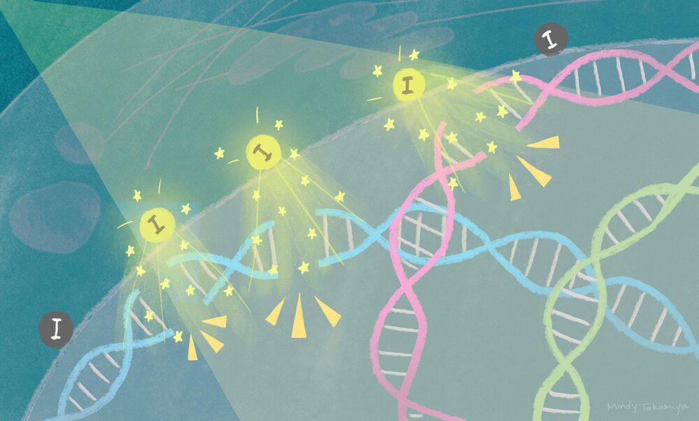
Researchers have found a way to enhance radiation therapy using novel iodine nanoparticles.
Cancer cell death is triggered within three days when X-rays are shone onto tumor tissue containing iodine-carrying nanoparticles. The iodine releases electrons that break the tumor’s DNA, leading to cell death. The findings, by scientists at Kyoto University’s Institute for Integrated Cell-Material Sciences (iCeMS) and colleagues in Japan and the US, were published in the journal Scientific Reports.
“Exposing a metal to light leads to the release of electrons, a phenomenon called the photoelectric effect. An explanation of this phenomenon by Albert Einstein in 1905 heralded the birth of quantum physics,” says iCeMS molecular biologist Fuyuhiko Tamanoi, who led the study. “Our research provides evidence that suggests it is possible to reproduce this effect inside cancer cells.”
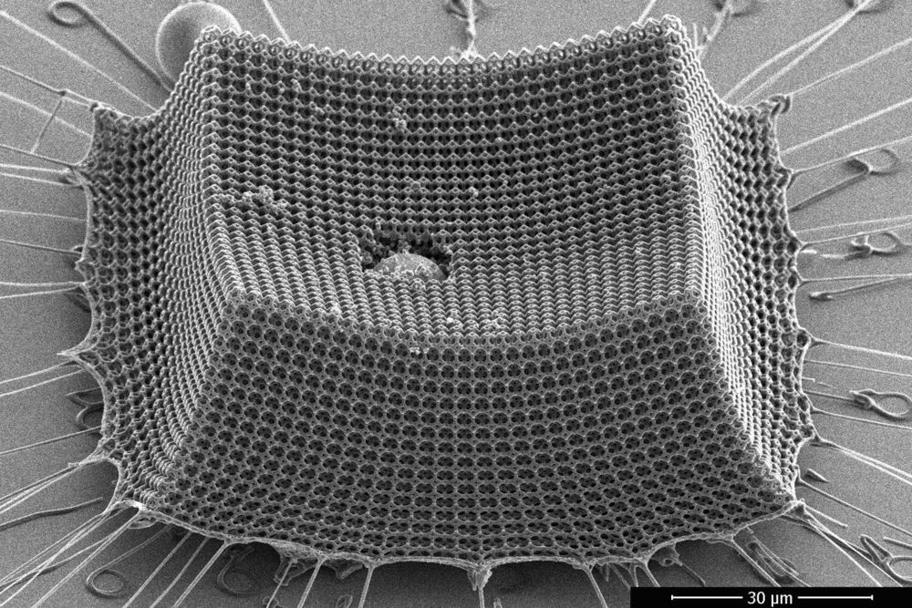
A new study by engineers at MIT, Caltech, and ETH Zürich shows that “nanoarchitected” materials—materials designed from precisely patterned nanoscale structures—may be a promising route to lightweight armor, protective coatings, blast shields, and other impact-resistant materials.
The researchers have fabricated an ultralight material made from nanometer-scale carbon struts that give the material toughness and mechanical robustness. The team tested the material’s resilience by shooting it with microparticles at supersonic speeds, and found that the material, which is thinner than the width of a human hair, prevented the miniature projectiles from tearing through it.
The researchers calculate that compared with steel, Kevlar, aluminum, and other impact-resistant materials of comparable weight, the new material is more efficient at absorbing impacts.
The innovative material that creates green energy through mechanical force.
A new nanotechnology development by an international research team led by Tel Aviv University researchers will make it possible to generate electric currents and voltage within the human body through the activation of various organs (mechanical force). The researchers explain that the development involves a new and very strong biological material, similar to collagen, which is non-toxic and causes no harm to the body’s tissues. The researchers believe that this new nanotechnology has many potential applications in medicine, including harvesting clean energy to operate devices implanted in the body (such as pacemakers) through the body’s natural movements, eliminating the need for batteries.
The study was led by Prof. Ehud Gazit of the Shmunis School of Biomedicine and Cancer Research at the Wise Faculty of Life Sciences, the Department of Materials Science and Engineering at the Fleischman Faculty of Engineering, and the Center for Nanoscience and Nanotechnology, along with his lab team, Dr. Santu Bera and Dr. Wei Ji.
Adding absorbent nanoparticles to polymer membranes simplifies desalination.
University of California, Berkeley, chemists have discovered a way to simplify the removal of toxic metals. like mercury and boron. during desalination to produce clean water, while at the same time potentially capturing valuable metals, such as gold.
Desalination — the removal of salt — is only one step in the process of producing drinkable water, or water for agriculture or industry, from ocean or waste water. Either before or after the removal of salt, the water often has to be treated to remove boron, which is toxic to plants, and heavy metals like arsenic and mercury, which are toxic to humans. Often, the process leaves behind a toxic brine that can be difficult to dispose of.