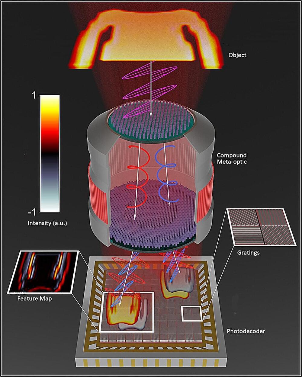
The device uses magnetic fields to carry out computation rather than shuttling electricity around, so it consumes far less power.
Category: nanotechnology – Page 134
By scientists from Inserm, CNRS, and Université de Montpellier at the Structural Biology Center in Montpellier. The nano-robot could lead to a closer study of the mechanical forces applied at microscopic levels, which are important for various biological and pathological processes.
The study was published in Nature Communications.
Cellular Mechanosensitivity
A novel method for measuring nanoparticle size could have applications in industry and basic materials science research.
Nanoparticles are present in everything from paints to pharmaceutical products. While nanoparticles have many important characteristics, such as molecular composition and shape, it is their size that determines many chemical and physical properties. A new technique relying on an optical vortex—a laser beam whose wave fronts twist around a dark central region—allows researchers to characterize nanoparticle size rapidly and continuously [1]. This light-based size probe might one day find applications in numerous industrial settings and aid fundamental materials science research.
It is difficult to precisely synthesize nanoparticles with the desired dimensions, so manufacturers must often validate that their nanoparticles have the right size to comply with regulations and to ensure product quality. There are many ways of determining nanoparticle size, but one popular approach, dynamic light scattering (DLS), is based on measurements of Brownian motion, the random particle movement caused by jostling from the surrounding liquid medium. In DLS, the Brownian motion is determined by measuring fluctuations in laser light scattering from the nanoparticles. In general, the faster the Brownian motion, the smaller the particles. But current techniques are generally not capable of characterizing the largest particles and measuring them continuously.
Flexible electronics have enabled the design of sensors, actuators, microfluidics and electronics on flexible, conformal and/or stretchable sublayers for wearable, implantable or ingestible applications. However, these devices have very different mechanical and biological properties when compared to human tissue and thus cannot be integrated with the human body.
A team of researchers at Texas A&M University has developed a new class of biomaterial inks that mimic native characteristics of highly conductive human tissue, much like skin, which are essential for the ink to be used in 3D printing.
This biomaterial ink leverages a new class of 2D nanomaterials known as molybdenum disulfide (MoS2). The thin-layered structure of MoS2 contains defect centers to make it chemically active and, combined with modified gelatin to obtain a flexible hydrogel, comparable to the structure of Jell-O.
Computer chip designers, materials scientists, biologists and other scientists now have an unprecedented level of access to the world of nanoscale materials thanks to 3D visualization software that connects directly to an electron microscope, enabling researchers to see and manipulate 3D visualizations of nanomaterials in real time.
Developed by a University of Michigan-led team of engineers and software developers, the capabilities are included in a new beta version of tomviz, an open-source 3D data visualization tool that’s already used by tens of thousands of researchers. The new version reinvents the visualization process, making it possible to go from microscope samples to 3D visualizations in minutes instead of days.
In addition to generating results more quickly, the new capabilities enable researchers to see and manipulate 3D visualizations during an ongoing experiment. That could dramatically speed research in fields like microprocessors, electric vehicle batteries, lightweight materials and many others.
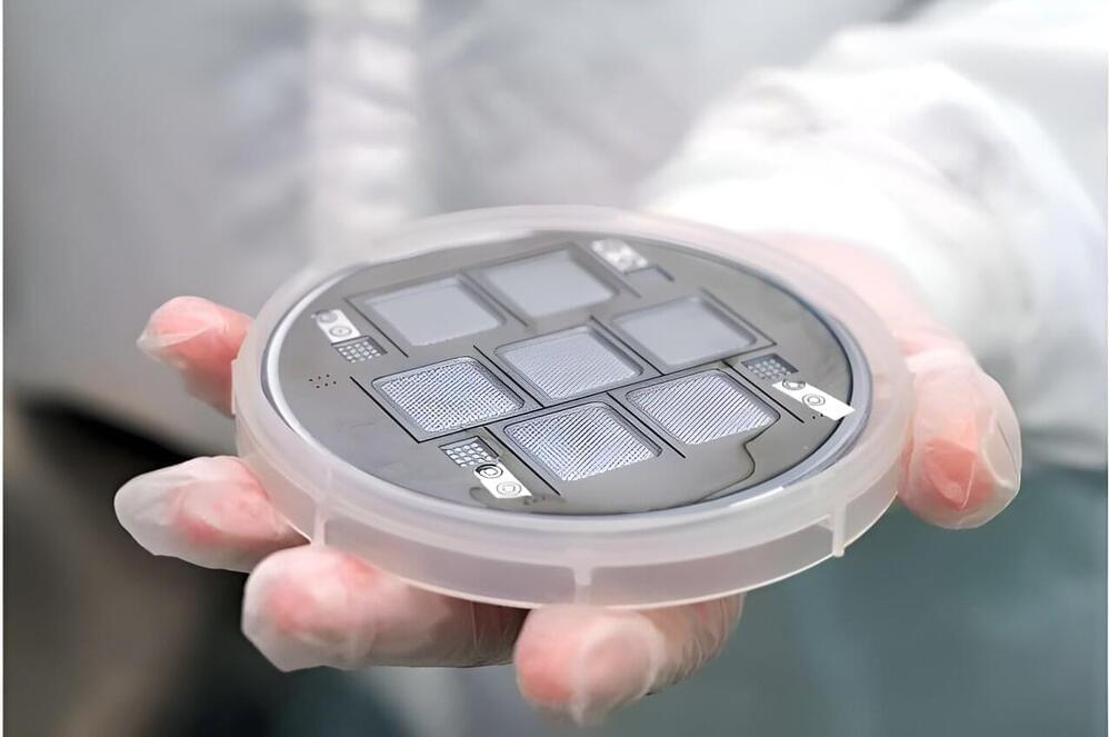
Technology on the nanometre scale could provide solutions to move on from the solid-state era.
Although chemotherapy can be a lifesaving treatment for patients with cancer, some of these medications can damage the heart. A team led by researchers at Massachusetts General Hospital (MGH) recently developed a nanoparticle probe that can detect an indicator of heart damage from chemotherapy.
Experiments with the probe also revealed that in mice with cancer, intermittent fasting before chemotherapy can prevent this heart damage indicator from arising, leading to preserved cardiac function and prolonged survival.
The study, which is published in Nature Biomedical Engineering, focused on autophagy—a process that cells use to remove unnecessary or dysfunctional components. A delicate balance exists between the protective and deleterious effects of this process: reduced levels of autophagy have been implicated in cardiovascular disease and other conditions; however, autophagy can also be a primary mechanism of cell death.
Circa 2012 o.o!!!
We report on two extensions of the traditional analysis of low-dimensional structures in terms of low-dimensional quantum mechanics. On one hand, we discuss the impact of thermodynamics in one or two dimensions on the behavior of fermions in low-dimensional systems. On the other hand, we use both quantum wells and interfaces with different effective electron or hole mass to study the question when charge carriers in interfaces or layers exhibit two-dimensional or three-dimensional behavior.
Most of us have felt the shock from static electricity by touching a metallic object after putting on a sweater or walking across a carpet. This occurs as a result of charge build-up whenever two dissimilar materials (such as our body and the fabric) come in contact with each other.
In 2012, scientists from the U.S. and China used this phenomenon, known as “triboelectric effect,” to build a triboelectric nanogenerator (TENG) that converts unused mechanical energy into useful electrical energy. Their device consisted of two triboelectric polymer films with metallic electrodes, which, when brought together and separated, resulted in charge separation and the development of an electric voltage sufficient to power small electronic devices.
Viewed as potential sustainable energy harvesters, efforts have been made to enhance the power output of TENGs by injecting charges to the surface of triboelectric films. However, charge recombination in the electrode and charge repulsion on the surface of the material prevents them from achieving high surface charge densities.
In recent years, ribonucleic acid (RNA) has emerged as a powerful tool for the development of novel therapies. RNA is used to copy genetic information contained in our hereditary material, the deoxyribonucleic acid (DNA), and then serves as a template for building proteins, the building blocks of life. Delivery of RNA into cells remains a major challenge for the development of novel therapies across a broad range of diseases. Researchers at the Max Planck Institute of Molecular Cell Biology and Genetics (MPI-CBG) in Dresden together with researchers from the global biopharmaceutical company AstraZeneca have investigated where and how mRNA is delivered inside the cell. They found that mRNA uses an unexpected entry door. Their results provide novel insights into the development of RNA therapeutics towards efficient delivery and lower dosages.
DNA (deoxyribonucleic acid) contains the genetic information required for the development and maintenance of life. This information is communicated by messenger ribonucleic acid (mRNA) to make proteins. mRNA-based therapeutics have the potential to address unmet needs for a wide variety of diseases, including cancer and cardiovascular disease. mRNA can be delivered to cells to trigger the production, degradation or modification of a target protein, something impossible with other approaches. A key challenge with this modality is being able to deliver the mRNA inside the cell so that it can be translated to make a protein. mRNA can be packed into lipid nanoparticles (LNPs)—small bubbles of fat—that protect the mRNA and shuttle it into cells. However, this process is not simple, because the mRNA has to pass the membrane before it can reach its site of action in the cell interior, the cytoplasm.
Researchers in the team of MPI-CBG director Marino Zerial are experts in visualizing the cellular entry routes of molecules in the cell, such as mRNA with high-resolution microscopes. They teamed up with scientists from AstraZeneca who provided the researchers with lipid nanoparticle prototypes that they had developed for therapeutic approaches to follow the mRNA inside the cell. The study is published in the Journal of Cell Biology.