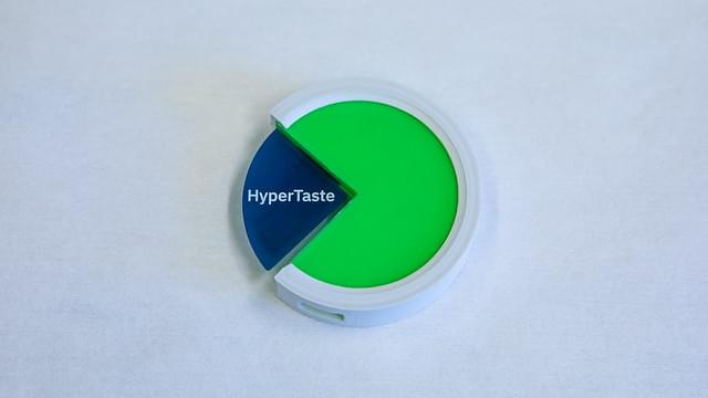
The tongue, called Hypertaste, can detect and analyze a liquid’s chemical composition.
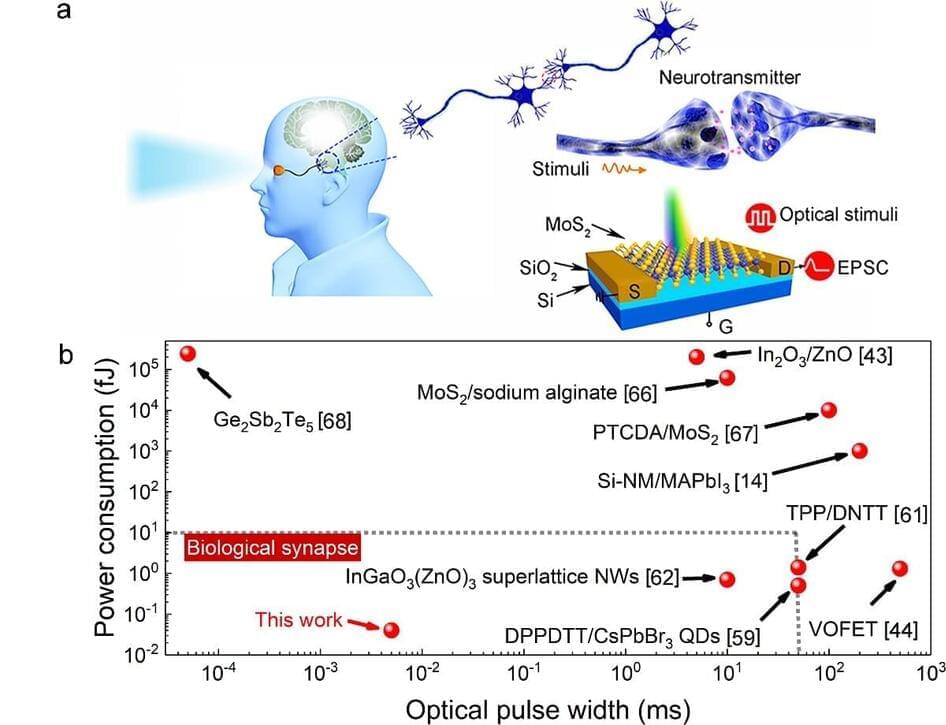
Neuromorphic photonics/electronics is the future of ultralow energy intelligent computing and artificial intelligence (AI). In recent years, inspired by the human brain, artificial neuromorphic devices have attracted extensive attention, especially in simulating visual perception and memory storage. Because of its advantages of high bandwidth, high interference immunity, ultrafast signal transmission and lower energy consumption, neuromorphic photonic devices are expected to realize real-time response to input data. In addition, photonic synapses can realize non-contact writing strategy, which contributes to the development of wireless communication.
The use of low-dimensional materials provides an opportunity to develop complex brain-like systems and low-power memory logic computers. For example, large-scale, uniform and reproducible transition metal dichalcogenides (TMDs) show great potential for miniaturization and low-power biomimetic device applications due to their excellent charge-trapping properties and compatibility with traditional CMOS processes. The von Neumann architecture with discrete memory and processor leads to high power consumption and low efficiency of traditional computing. Therefore, the sensor-memory fusion or sensor-memory-processor integration neuromorphic architecture system can meet the increasingly developing demands of big data and AI for low power consumption and high performance devices. Artificial synaptic devices are the most important components of neuromorphic systems. The performance evaluation of synaptic devices will help to further apply them to more complex artificial neural networks (ANN).
Chemical vapor deposition (CVD)-grown TMDs inevitably introduce defects or impurities, showed a persistent photoconductivity (PPC) effect. TMDs photonic synapses integrating synaptic properties and optical detection capabilities show great advantages in neuromorphic systems for low-power visual information perception and processing as well as brain memory.
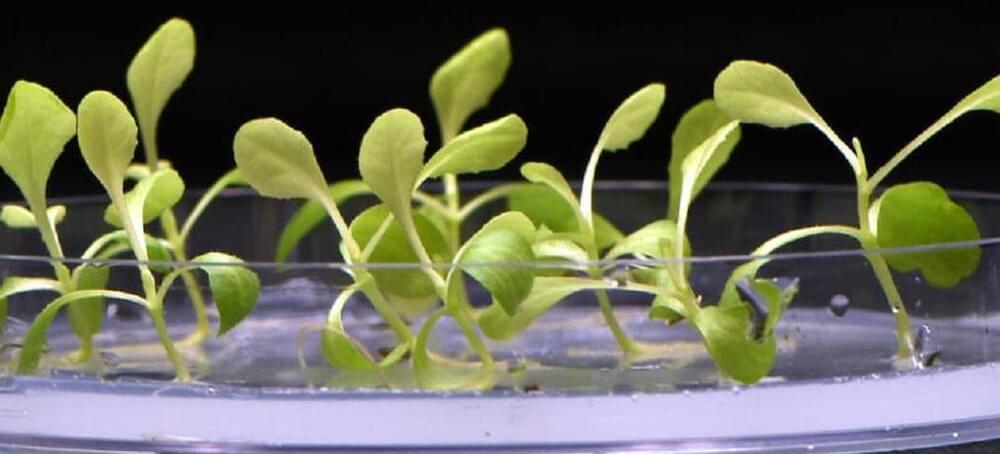
Photosynthesis uses a series of chemical reactions to convert carbon dioxide, water, and sunlight into glucose and oxygen. The light-dependent stage comes first, and relies on sunlight to transfer energy to plants, which convert it to chemical energy. The light-independent stage (also called the Calvin Cycle) follows, when this chemical energy and carbon dioxide are used to form carbohydrate molecules (like glucose).
A research team from UC Riverside and the University of Delaware found a way to leapfrog over the light-dependent stage entirely, providing plants with the chemical energy they need to complete the Calvin Cycle in total darkness. They used an electrolysis to convert carbon dioxide and water into acetate, a salt or ester form of acetic acid and a common building block for biosynthesis (it’s also the main component of vinegar). The team fed the acetate to plants in the dark, finding they were able to use it as they would have used the chemical energy they’d get from sunlight.
They tried their method on several varieties of plants and measured the differences in growth efficiency as compared to regular photosynthesis. Green algae grew four times more efficiently, while yeast saw an 18-fold improvement.
Hear from Nobel laureate Jennifer Doudna on the four ways that CRISPR gene editing technologies will revolutionize healthcare.
In her 31 March talk at the Frontiers Forum, Prof Jennifer Doudna outlined how CRISPR-based therapies are already transforming the lives of patients with previously limited treatment options. She also gave her vision for how her serendipitous discovery will revolutionize healthcare for us all. The session was attended by over 9,200 representatives from science, policy and business across the world.
Jennifer’s keynote talk was followed by a discussion with global experts on access and ethical considerations:
• Prof Andrea Crisanti, Imperial College London.
• Prof Françoise Baylis, Dalhousie University.
• Dr Soumya Swaminathan, Chief Scientist, World Health Organization.
2022 marks the 10th anniversary of Jennifer’s groundbreaking development of CRISPR-Cas9 as a genome-engineering technology, with collaborator Prof Emmanuelle Charpentier. The two earned the 2020 Nobel Prize in Chemistry for their work, which has forever changed the course of human and agricultural genomics research. Jennifer Doudna is the Li Ka Shing Chancellor’s Chair and a Professor in the Departments of Chemistry and of Molecular and Cell Biology at the University of California, Berkeley, and Founder of the Innovative Genomics Institute.
The Frontiers Forum showcases science-led solutions for healthy lives on a healthy planet. Watch previous sessions at https://forum.frontiersin.org.
MAIN TALK
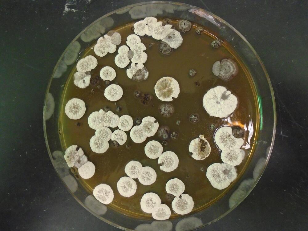
Aircrafts transport people, ship goods, and perform military operations, but the petroleum-based fuels that power them are in short supply. In research publishing on June 30 in the journal Joule, researchers at the Lawrence Berkeley Lab have found a way to generate an alternative jet fuel by harvesting an unusual carbon molecule produced by the metabolic process of bacteria commonly found in soil.
“In chemistry, everything that requires energy to make will release energy when it’s broken,” says lead author Pablo Cruz-Morales, a microbiologist at DTU Biosustain, part of the Technical University of Denmark. When petroleum jet fuel is ignited, it releases a tremendous amount of energy, and the scientists at the Keasling Lab at the Lawrence Berkeley Laboratory thought there must be a way to replicate this without waiting millions of years for new fossil fuels to form.
Jay Keasling, a chemical engineer at University of California, Berkeley, approached Cruz-Morales, who was a postdoc in his lab at the time, to see if he could synthesize a tricky molecule that has the potential to produce a lot of energy. “Keasling told me: it’s gonna be an explosive idea,” says Cruz-Morales.
Researchers at North Carolina State University show that an important gene in maize called HPC1 modulates certain chemical processes that contribute to flowering time, and has its origins in “teosinte mexicana,” a precursor to modern-day corn that grows wild in the highlands of Mexico. The findings provide insight into plant evolution and trait selection, and could have implications for corn and other crops’ adaptation to low temperatures.
“We are broadly interested in understanding how natural variation of lipids are involved in the growth and development of plants, and how these compounds may help plants adapt to their immediate environments,” said Rubén Rellán-Álvarez, assistant professor of structural and molecular biochemistry at NC State and the corresponding author of a paper describing the research. “Specifically, we wanted to learn more about variation in lipids called phospholipids, which consist of phosphorus and fatty acids, and their role in adaptation to cold, low phosphorus, and the regulation of important processes for plant fitness and yield like flowering time.”
Maize grown at higher altitudes, like the highlands of Mexico, needs special accommodations in order to grow successfully. The colder temperatures in these mountainous regions put maize at a slight disadvantage when compared with maize grown at lower elevations and higher temperatures.
In the future, a woman with a spinal cord injury could make a full recovery; a baby with a weak heart could pump his own blood. How close are we today to the bold promise of bionics—and could this technology be used to improve normal human functions, as well as to repair us? Join Bill Blakemore, John Donoghue, Jennifer French, Joseph J. Fins, and P. Hunter Peckham at “Better, Stronger, Faster,” part of the Big Ideas Series, as they explore the unfolding future of embedded technology.
This program is part of the Big Ideas Series, made possible with support from the John Templeton Foundation.
Visit our Website: http://www.worldsciencefestival.com/
Like us on Facebook: https://www.facebook.com/worldscience… us on twitter: https://twitter.com/WorldSciFest Original Program date: May 31, 2014 Host: Bill Blakemore Participants: John Donoghue, Jennifer French, Joseph J. Fins, P. Hunter Peckham Re-engineering the anatomy of the “Vitruvian Man” 00:00 Bill Blakemore’s Introduction. 2:06 Participant introductions. 4:27 What is FES? (Functional Electrical Stimulation) 6:06 A demonstration with FES and without. 10:06 How did you test FES systems? 14:16 Jen French the first bionic pioneer. 16:40 What was the journey like from injury to today? 18:35 A live demonstration of FES. 20:40 What is BrainGate? 27:55 What is the potential for this technology? 37:00 When will this technology be publicly available? 40:50 A cell phone app to drink water or stand up? 44:55 Jen French would be the first to try new technology. 50:39 What is the history of altering the human brain? 1:00:57 The move from chemical to electrical medical care. 1:05:40 The challenge of what is going to drive the delivery of care to groups in need. 1:11:36 Can these devices be implanted without surgery? 1:18:13 What field needs the most funding for this to become available to everyone? 1:19:40 What are the numbers of people who can use this technology? 1:23:44 Why can’t we use stem cells to reconnect human spinal tissue? 1:25:37 What is the collaboration level between institutions? 1:29:16 How far away are we from using brain waves to control objects and communicate with each other? 1:30:20
Follow us on twitter: https://twitter.com/WorldSciFest.
Original Program date: May 31, 2014
Host: Bill Blakemore.
Participants: John Donoghue, Jennifer French, Joseph J. Fins, P. Hunter Peckham.
Re-engineering the anatomy of the “Vitruvian Man” 00:00.
Bill Blakemore’s Introduction. 2:06
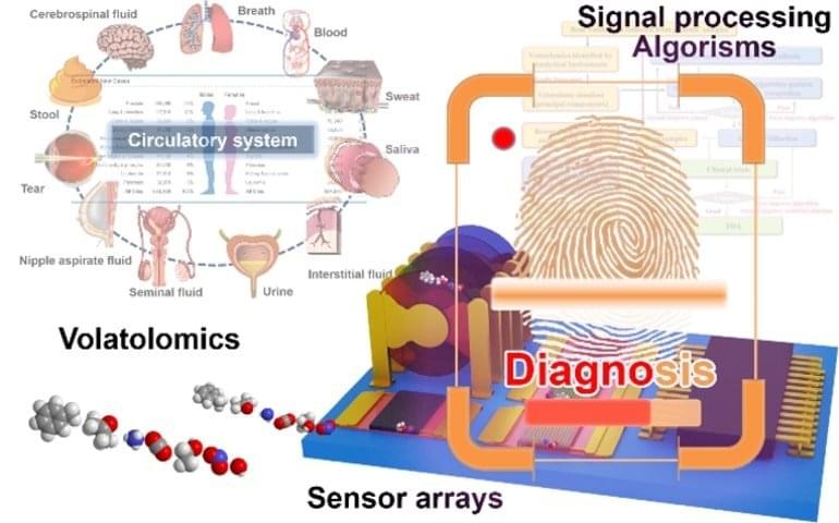
Summary: A new robotic system can identify volatile organic compounds associated with diseases by analyzing bodily emissions.
Source: Tsinghua University Press.
Scientists are working on diagnostic techniques that could sniff out chemical compounds from breath, sweat, tears and other bodily emissions and that act as fingerprints of thousands of diseases.
The theory, design, and operation of a nuclear propulsion engine advantages are explained verses conventional chemical rockets such as the Saturn V.

Blockchain is a digital technology that allows a secure and decentralized record of transactions that is increasingly used for everything from cryptocurrencies to artwork. But Yale researchers have found a new use for blockchain: they’ve leveraged the technology to give individuals control of their own genomes.
Their findings are published June 29 in the journal Genome Biology.
“Our primary goal is to give ownership of genomic data back to the individual,” said senior author Mark Gerstein, the Albert L. Williams Professor of Biomedical Informatics and professor of molecular biophysics and biochemistry, of computer science, and of statistics and data science.