A new study has revealed the clearest-ever picture of the surface chemistry of worm species that provides insights into how animals interact with their environment and each other. These discoveries could pave the way for strategies to deepen our understanding of evolutionary adaptations, refine behavioral research, and ultimately overcome parasitic infections.
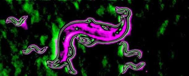
Scientists from the University’s School of Pharmacy used an advanced mass spectrometry imaging system to examine the nematodes Caenorhabditis elegans and Pristionchus pacificus, aiming to characterize species-specific surface chemical composition and its roles in physiology and behavior.
Their results show that nematode surfaces are predominantly oily or lipid-based, forming a complex chemical landscape. The findings have been published in theJournal of the American Chemical Society.