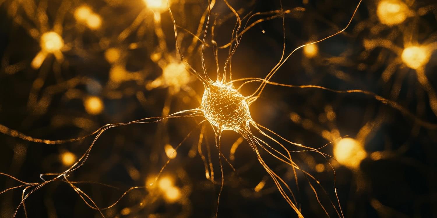
To study what happened in the brain during this task, the researchers used functional magnetic resonance imaging, which measures blood flow as a proxy for neural activity. They scanned the brains of 11 participants while they performed the memory task over multiple sessions. By applying a complex decoding model to the imaging data, the researchers were able to estimate not only what participants were remembering but also how uncertain they were about each memory. The model treated neural activity as a probabilistic code, where stronger or more focused patterns of activity reflected more confident memory representations.
The results showed that neural signals in the visual cortex—the area of the brain involved in processing visual information—were more intense for the high-priority memory items. These stronger signals translated to smaller memory errors and greater confidence. On average, participants remembered the high-priority items more accurately and responded more quickly when asked to recall them. Their eye movements were closer to the correct location, and they took less time to respond. These behavioral improvements matched the patterns observed in the brain data.
The study also found that the magnitude of neural activity in the frontal cortex predicted how well participants could distinguish between high-and low-priority memories. This suggests that the frontal cortex plays a regulatory role, sending signals that adjust the strength of memory representations in visual areas depending on how important each item is. In other words, the frontal brain regions help direct the mental spotlight, increasing the “volume” of the memories that matter most.


