A new single-protein analysis technique gives researchers an unprecedented ability to study proteins called scramblases, which have critical roles in biology. The development of the new technique, in a study led by investigators at Weill Cornell Medicine and Ruhr University Bochum in Germany, expands the toolkit available to cell biologists and biophysicists and could someday be useful in devising new strategies against multiple diseases.
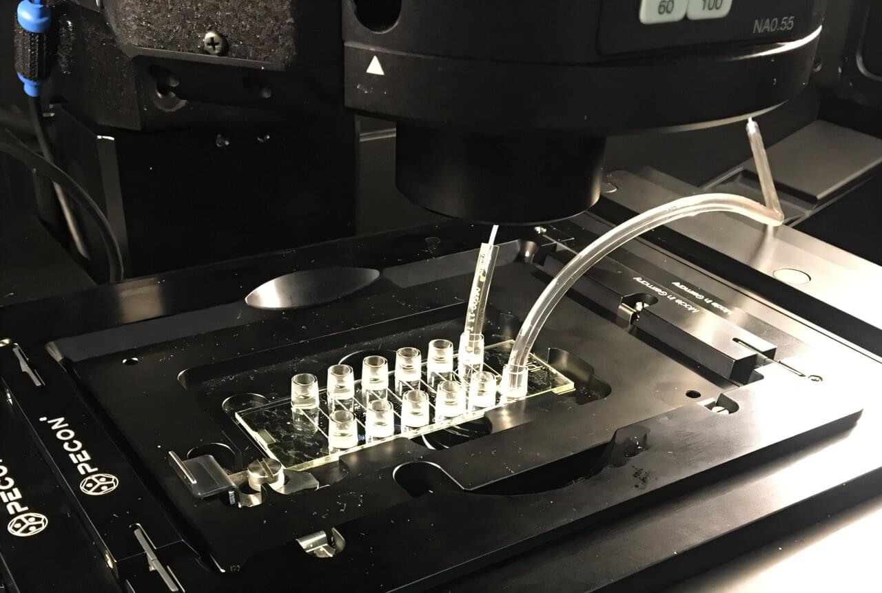
Scramblases operate within cell membranes to rearrange the fat-related molecules, known as lipids, that make up those membranes. Their disruption of the usual layered organization of the membrane is essential for many important biological processes. In the study, published in Nature Structural & Molecular Biology, the researchers developed a fluorescence imaging-based technique—the first of its kind—for measuring the activity rates of individual scramblase proteins. Their demonstrations of the technique uncovered new findings on key scramblases and showcased the technique’s broad applicability.
“I’m excited about this new platform as it is versatile and provides unprecedented information on exactly how fast a single scramblase works,” said study co-senior author Dr. Anant Menon, professor of biochemistry and biophysics at Weill Cornell Medicine.