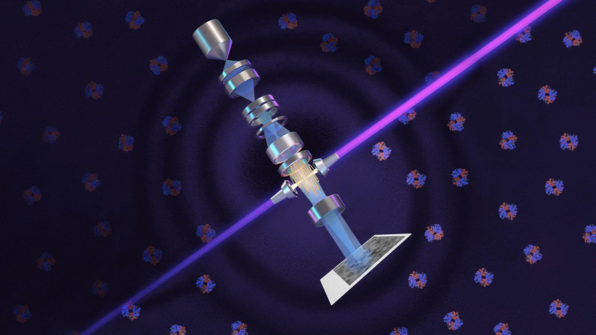
Researchers in the United States have built a technology that boosts the performance of electron microscopes. Berkeley Lab and UC Berkeley physicists’ new technique offers detailed images of the small molecules and cell structures that are crucial to understanding biology and disease.
They have adapted the phase-contrast technique to cryo-electron microscopy (cryo-EM), which has about 10,000 times the magnification of light microscopy. Their laser-based phase plate produces sharp images of molecules that today’s cutting-edge cryo-EM systems struggle to capture.
The research team revealed that the new technology was brought to fruition by more than 15 years of theoretical and experimental work by leading microscopy scientists, collaboration with expert machinists, and support from Biohub.