Nearly 100 years ago, a seemingly simple discovery revolutionized the microscope. The introduction of phase contrast, which garnered a Nobel Prize in 1953, brought into clear view structures inside cells that had previously been too faint or washed out for biologists to study.
UC Berkeley physicists have now adapted the phase-contrast technique to the electron microscope, which has about 10,000 times the magnification of microscopes using optical light. The study is published in the journal Science.
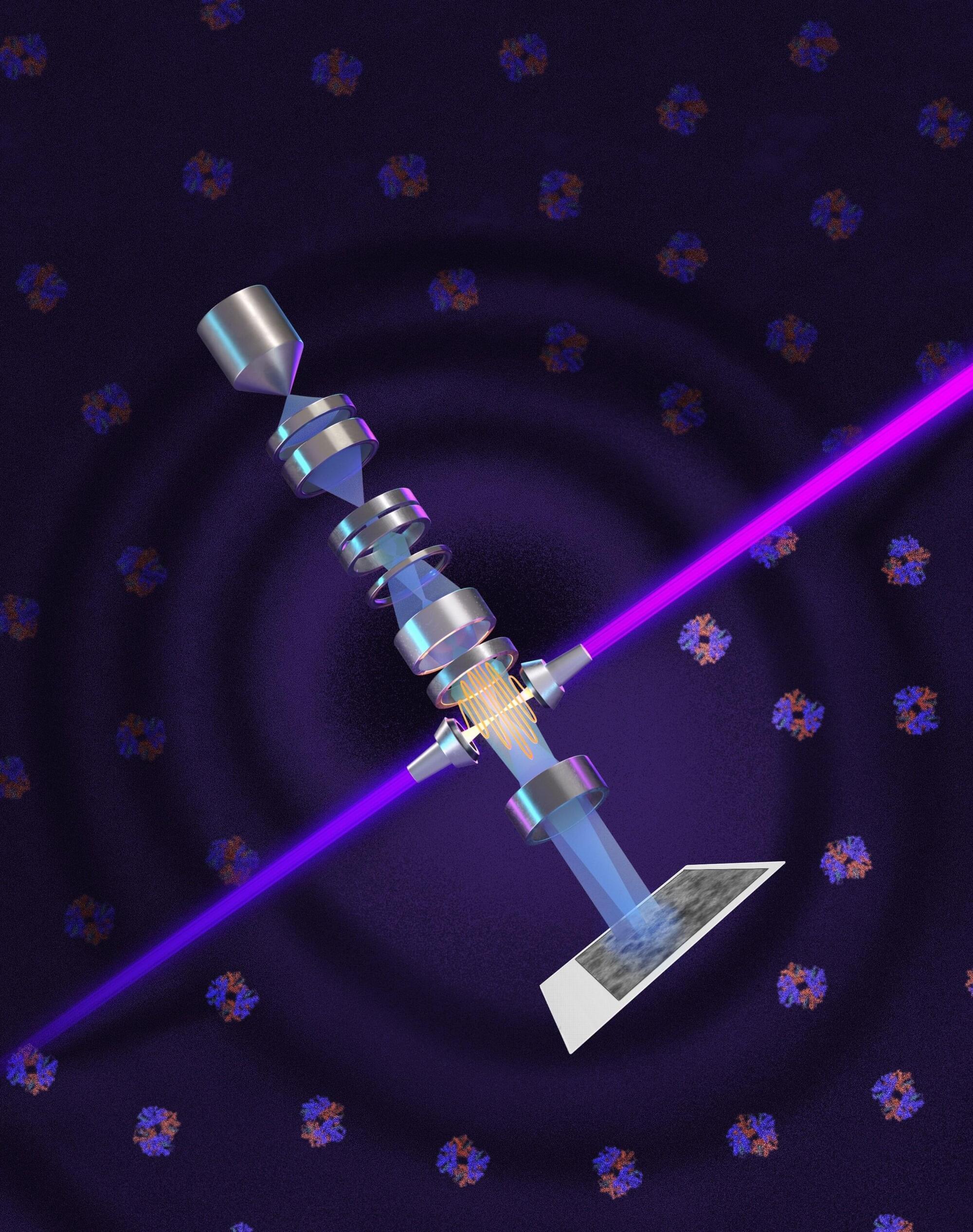
The addition of a so-called laser phase plate has the potential to greatly improve cryoelectron microscopy (cryo-EM), a technique for determining the structure of molecules that itself revolutionized the understanding of proteins and accelerated new drug discovery starting a decade ago.