FNDC5/irisin detection was performed by a sandwich ELISA reaction using DuoSet® ELISA Development Systems kit (R&D Systems), according to manufacturer’s instructions. Blood samples were collected at 10 or 40 dpi, allowed to sit for at least 1 h, and then centrifuged for 10 min at 224 g in a refrigerated centrifuge (4 °C) to isolate the serum. Samples were stored at −80 °C until irisin detection.
Serum samples were assayed for TNF-α, INF-γ, IL-2, IL-4, IL-6, IL-10, and IL-17a using a Cytometric Bead Array (CBA) Th1/ Th2/ Th17 kit (BD Biosciences), according to the manufacturer’s instructions. Data were acquired using a Cytoflex S (Beckman Coulter) flow cytometer. After data acquisition, dedicated software (FCAP Array, BD Biosciences) was used to analyze the results by gating bead populations, calculating MFI values, generating standard curves, and determining analyte concentrations. These analyses were performed at the Flow Cytometry Facility of the Instituto Oswaldo Cruz (Fiocruz).
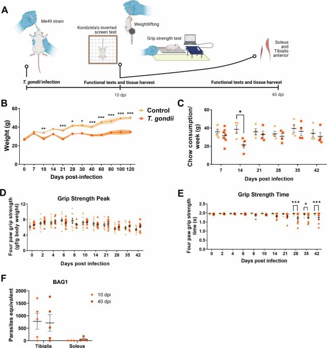
T. gondii infection was quantified using RT-qPCR with bag1 and enolase2 primers to detect bradyzoite and tachyzoite forms, respectively, according to49. Ct values were compared to a standard curve amplification, derived from known T. gondii RNA concentrations. The standard curve was constructed with six 10-fold dilutions, starting with 6.0 × 10⁶ parasites for either bradyzoites or tachyzoites. Primer sequences are available in Table 2.