To understand what drives disease progression in tissues, scientists need more than just a snapshot of cells in isolation—they need to see where the cells are, how they interact, and how that spatial organization shifts across disease states. A computational method called MESA (Multiomics and Ecological Spatial Analysis), detailed in a study published in Nature Genetics, is helping researchers study diseased tissues in more meaningful ways.
The work details the results of a collaboration among researchers from MIT, Stanford University, Weill Cornell Medicine, the Ragon Institute of MGH, MIT, and Harvard, and the Broad Institute of MIT and Harvard, and was led by the Stanford team.
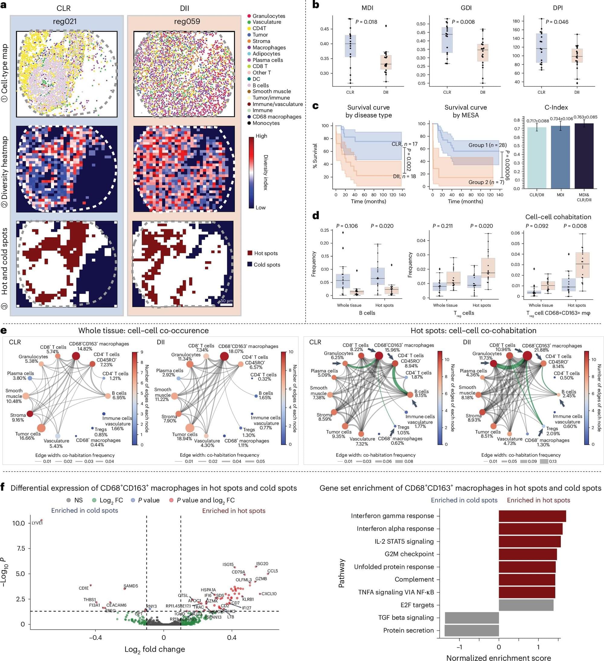
MESA brings an ecology-inspired lens to tissue analysis. It offers a pipeline to interpret spatial omics data—the product of cutting-edge technology that captures molecular information along with the location of cells in tissue samples. This data provides a high-resolution map of tissue “neighborhoods,” and MESA helps make sense of the structure of that map.