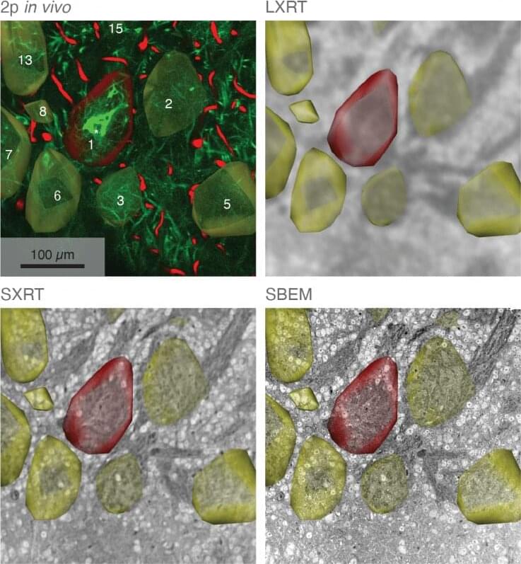
Researchers at the Francis Crick Institute have developed an imaging technique to capture information about the structure and function of brain tissue at subcellular level—a few billionths of a meter, while also capturing information about the surrounding environment.
The unique approach detailed in Nature Communications today (25 May), overcomes the challenges of imaging tissues at different scales, allowing scientists to see the surrounding cells and how they function, so they can build a complete picture of neural networks in the brain.
Various imaging methods are used to capture information about tissue, cells and subcellular structures. However, a single method can only capture information about either the structure or function of the tissue and looking in detail at a nanometer scale means scientists lose information about the wider surroundings. This means that to gain an overall understanding of the tissue, imaging techniques need to be combined.
