
The immortal jellyfish is capable of winding back its lifecycle to become young again. Scientists are unlocking its cellular secrets to find ways human cells could rejuvenate too.
Get the latest international news and world events from around the world.

Railway Says Intruder May Have Accessed Customer Secrets, Urges Credential Rotation
Railway says an intruder may have accessed decrypted environment variables for some users, as a researcher reports related cryptocurrency theft.

Dynamic force sensing by bone delays aging and extends lifespan
Bai et al. show that bone acts as a dynamic force sensor that systemically delays aging and extends lifespan. This work reveals a bone-derived mechanism linking mechanical sensing to organismal longevity, suggesting new targets for anti-aging interventions.

Diabetes May Change Key Mitochondrial Signals in Alzheimer’s Disease
Researchers measured mitochondrial DNA copy number in post-mortem brain tissue. Alzheimer’s disease was associated with lower mtDNA levels, while diabetes was linked to higher levels.
Light-based method enables precise control of ultrafast electron flow in a scanning tunneling microscope
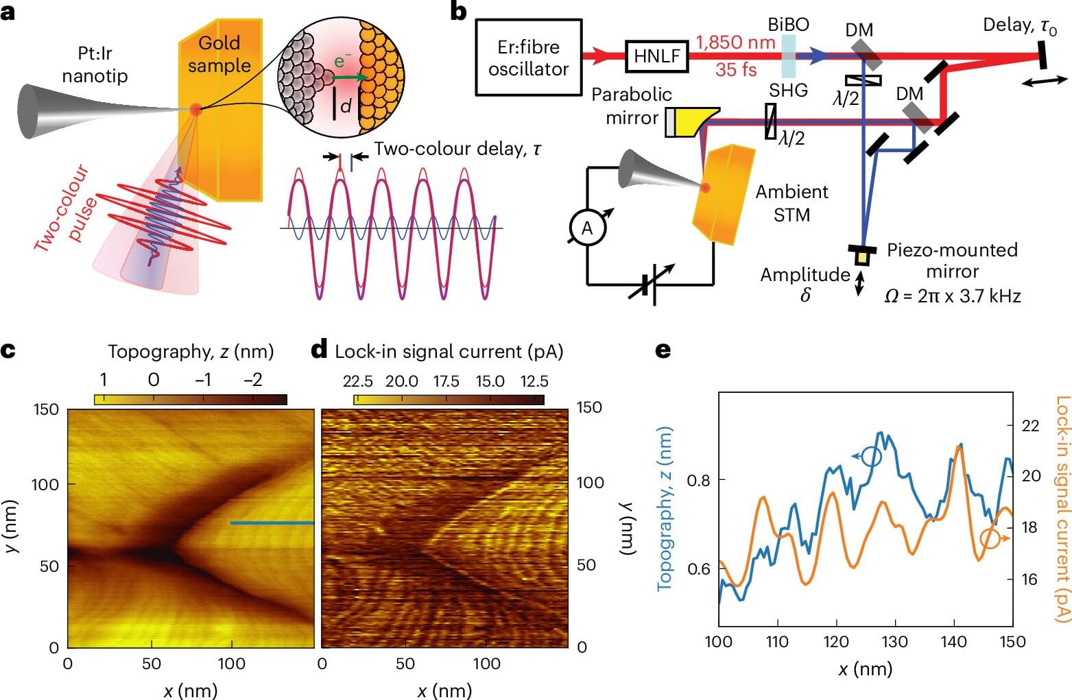
What would it take to watch an electron move through a material? Researchers would need to see individual atoms and capture an electron’s motion at incredibly short timescales. A Technion-led team has brought those two capabilities together in a scanning tunneling microscope, using carefully shaped laser pulses to control the direction of electron flow while preserving nanoscale spatial resolution.
The study, led by Michael Krüger of the Technion Faculty of Physics and published in Nature Photonics, builds on a microscope with an exceptionally sharp needle tip. When the needle tip is brought very close to a surface, electrons can cross the gap through a quantum effect called tunneling. The resulting current flow lets researchers map minute features of the surface. Until now, following the rapid movement of electrons within that tiny gap has been a major challenge.
The team directed infrared laser pulses into the gap between the tip and the sample. Each pulse contained two colors of light. By changing their relative timing, the researchers changed the shape of the light wave—and could steer electrons from the needle tip to the sample or back in the opposite direction. They adjusted the timing instead of repeatedly turning the laser on and off, helping them distinguish the ultrafast current from noise signals caused by laser heating.
ALMA spots ‘dragon egg’ that may represent the earliest stages of massive star formation
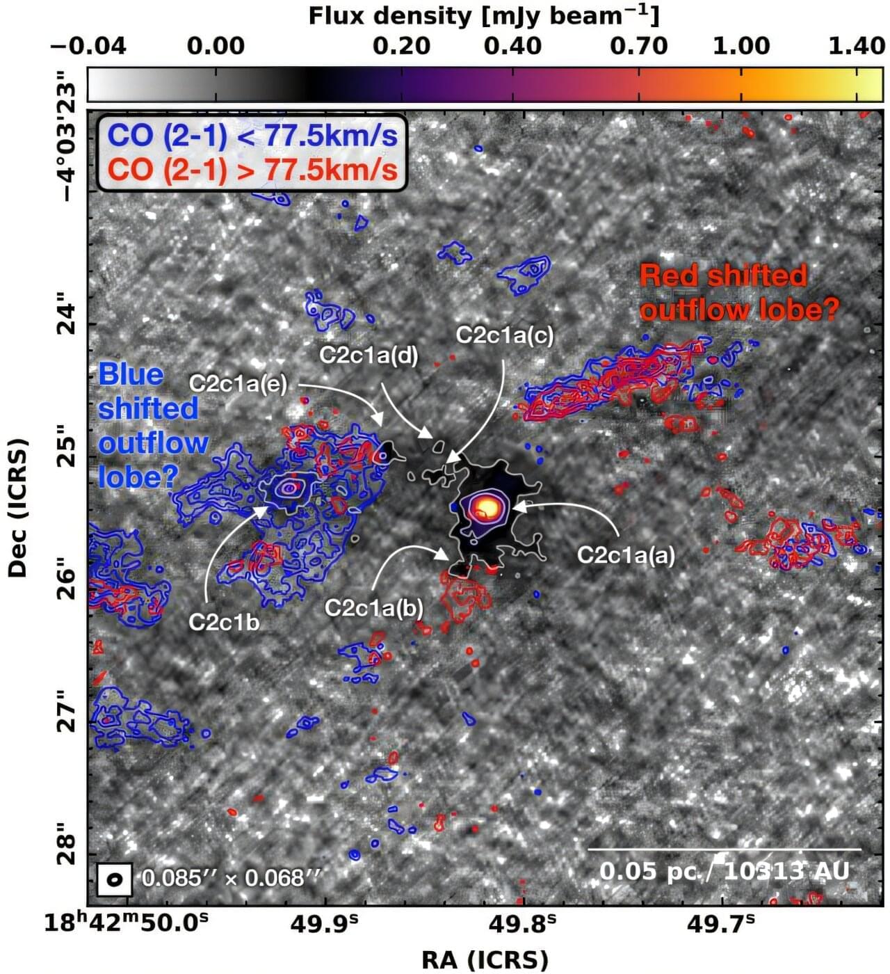
Astronomers identified an exceptionally tiny core that may represent an early stage in the formation of a massive star. Located in an infrared-dark area called Dragon Cloud, the core is nicknamed the “dragon egg.” The paper, accepted for publication in Astronomy & Astrophysics, was posted to the arXiv preprint server on Oct. 5.
How giant stars, with masses eight times greater than the sun, form is still a matter of debate in the scientific community. There are two main ideas. One says that a dense cloud of gas first condenses into a massive “pre-stellar” core, which then collapses to form a single giant star. Another says the star begins small and grows by pulling in material from the larger clump around it.
Researchers think that if they directly observe a compact, massive yet young concentration of mass, they will be able to lean toward one of these ideas. Such cores, however, are hard to detect, and if they are found, their interpretations are sometimes complicated by nearby star formation and stellar activity.
DNA programs a cell-free material to build its own motors and move
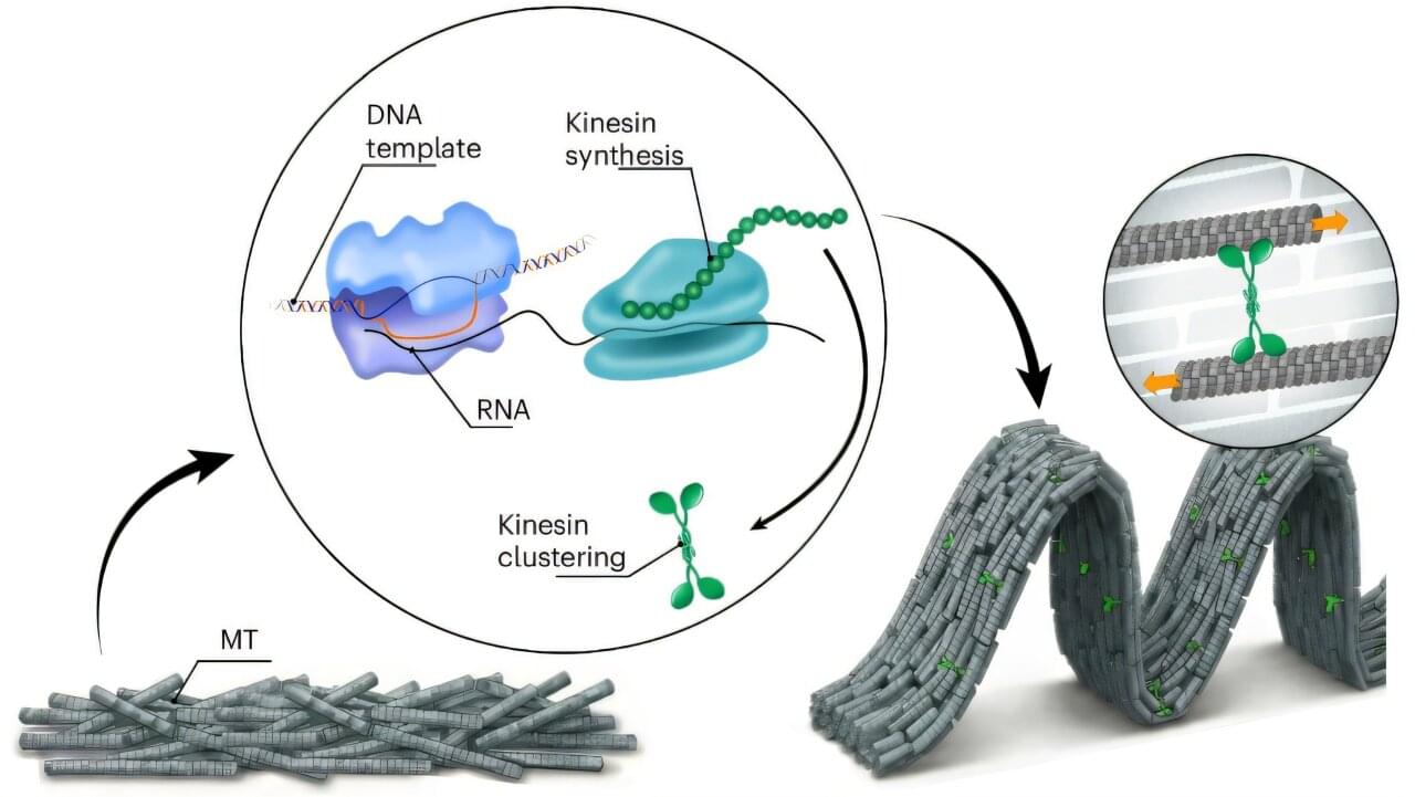
Researchers have created a material that manufactures the proteins responsible for cell movement, without needing intact living cells. By embedding cell-free gene expression into a thin sheet of protein filaments, they were able to make the material wrinkle, rise into blister-like shapes and break into bundles that flow around one another.
The instructions driving these transformations are built into the material itself—in DNA. Changing the genetic program made the filaments sustain flowing motion, stop after a burst of activity or repeatedly speed up and slow down.
The study, published in Nature Materials, demonstrates mechanical behavior that is “genetically programmed and executed autonomously,” as the authors describe it. Once assembled, the material can change its activity without researchers intervening at each step.
Experiment shows when quantum ‘cat’ states stop bouncing back
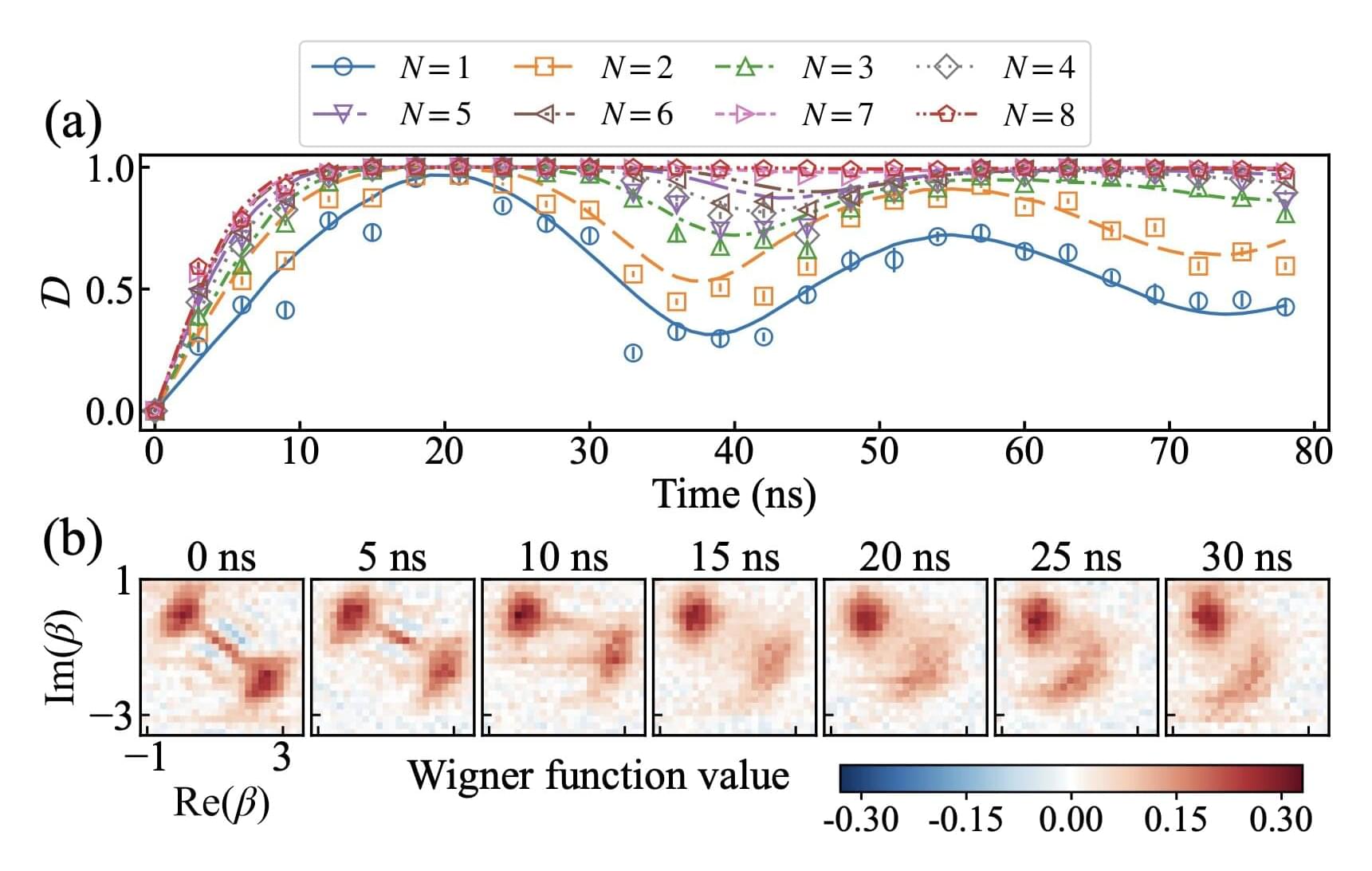
Quantum systems can occupy superpositions, quantum states that combine different possibilities. For instance, a microwave field can be prepared in a superposition of an empty state and a state that contains microwave light.
These two components can interfere, reinforcing or canceling each other’s contributions to the probabilities of measurement outcomes. This interference distinguishes a superposition from a situation in which the microwave field is simply empty or contains light.
A quantum system’s interactions with the surrounding environment can cause interference to fade, a process known as decoherence.
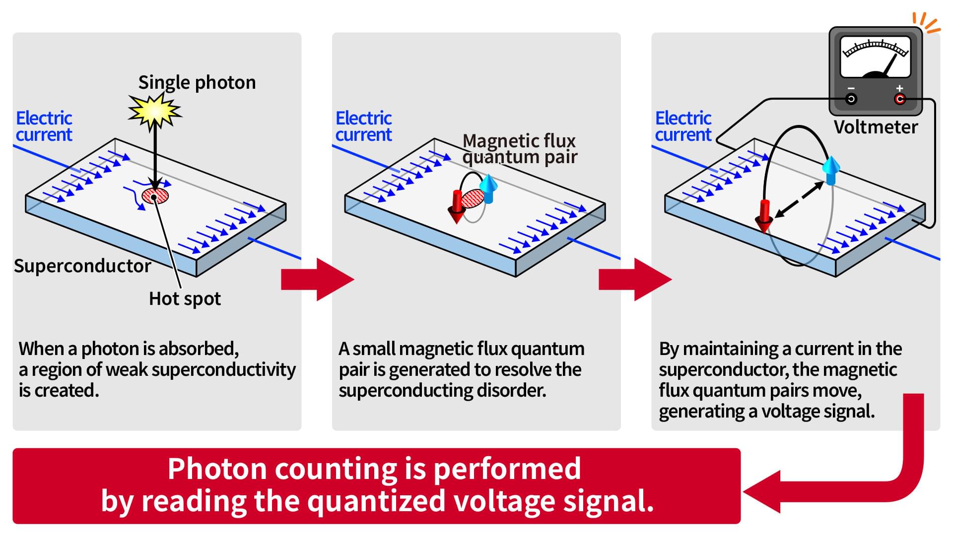
Counting magnetic flux quanta offers new way to measure photon numbers
Researchers at AIST have directly observed magnetic flux quanta generated in a superconductor by photon absorption and demonstrated a new principle of photon detection that identifies the number of photons based on the number of these flux quanta.
Detecting individual photons is a key technology for photonic quantum computers and quantum communication, and several methods have been proposed. While some of these methods are already in practical use for research purposes, their integration into industrial applications has just begun.
In a study appearing in the journal Physical Review Applied, a new approach to photon detection combines two important capabilities: high photon-number resolution, which is difficult to achieve with conventional technologies, and high-speed operation, which is desirable for industrial applications.
Chemically modified sugars enable direct nanostructure formation
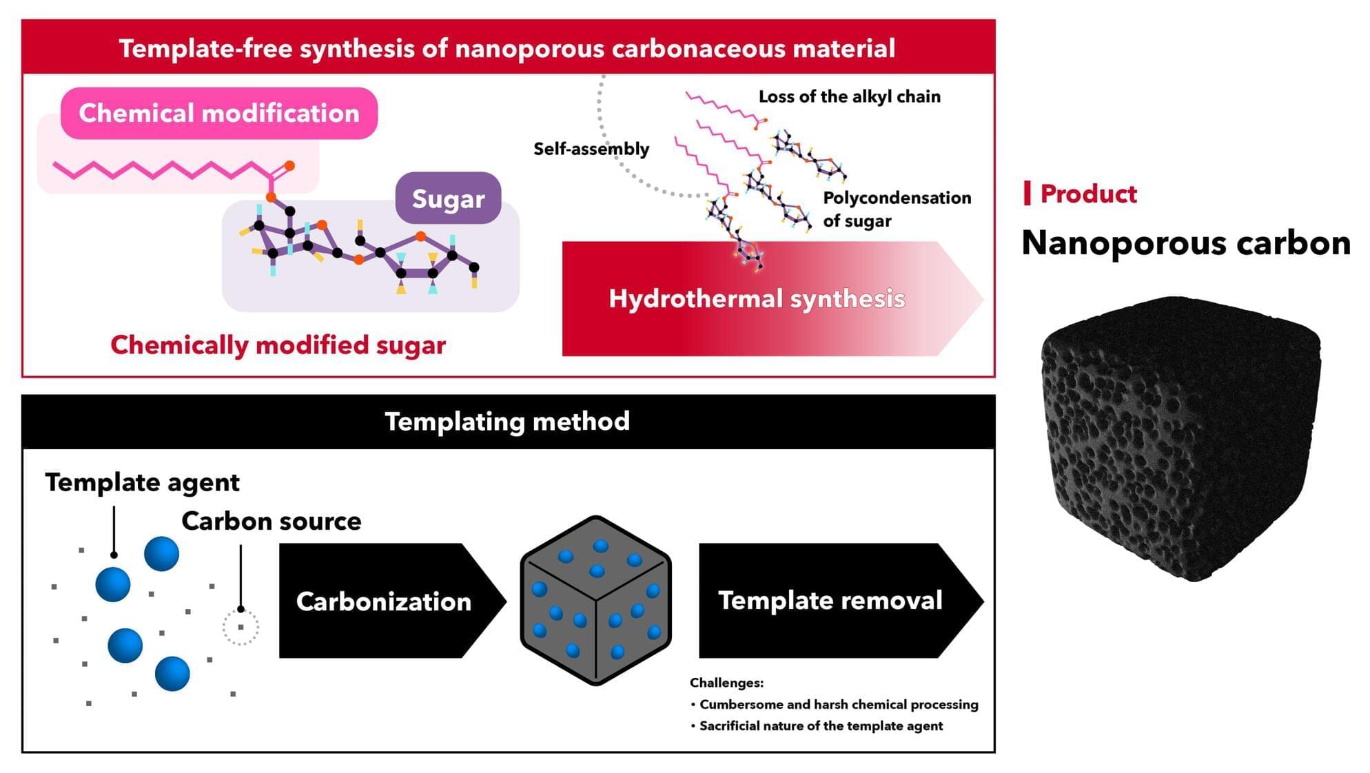
Researchers at AIST have demonstrated the synthesis of nanoporous carbonaceous materials and carbonaceous materials with layered nanostructures through a simple process involving the chemical modification of common sugars such as sucrose and fructose.
Nanoporous carbonaceous materials have attracted attention as materials that can help address energy-related challenges, with potential applications as catalyst supports and electrode materials for secondary batteries. In particular, carbonaceous materials with highly uniform pore sizes and shapes, which can be tailored for specific applications, are in increasing demand.
The conventional templating method, however, has several drawbacks, including the need for sacrificial template agents and complex synthesis procedures that consume resources and generate waste. Furthermore, some templating procedures rely on energy-intensive and harsh chemical processes involving high temperatures and strong acids.