PDF | On Jan 1, 2025, Vo Thi Thu Thao publishedFind, read and cite all the research you need on ResearchGate
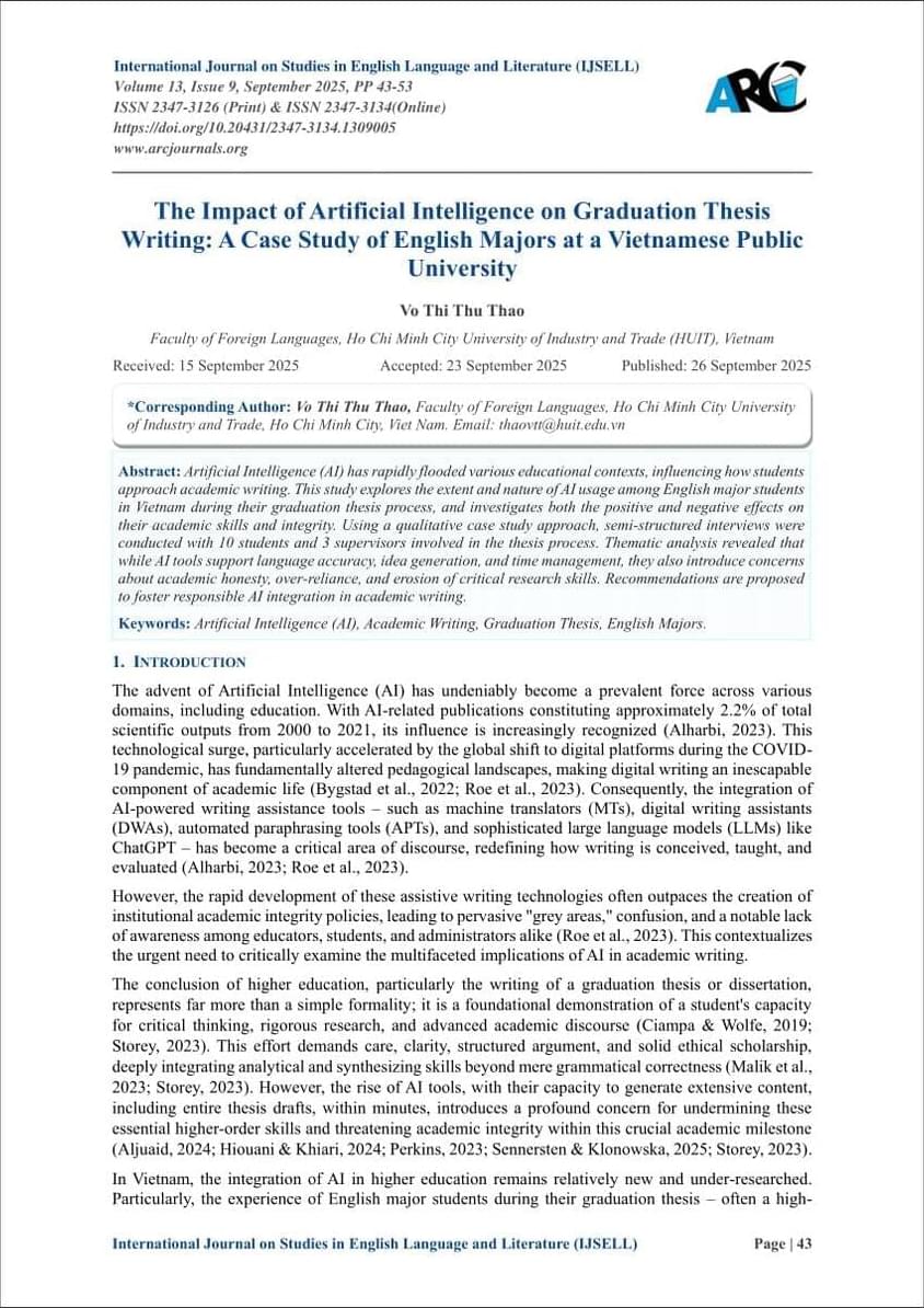
A KAIST research team has developed a next-generation world model, an internal model an AI builds to understand and predict the world, that learns executable theories from observation alone.
The team led by Professor Sungjin Ahn from the School of Computing proposed a new learning paradigm called Learning-to-Theorize (L2T), which trains AI to theorize how the world works using only observed information. The team also built the Neural Theorizer (NEO), a neural network-based model that implements this paradigm.
The research was presented at the 43rd International Conference on Machine Learning (ICML 2026), held in Seoul from July 6–11. The paper, published on the arXiv preprint server, was also selected for the Best Paper Award at the Compositional Learning Workshop.
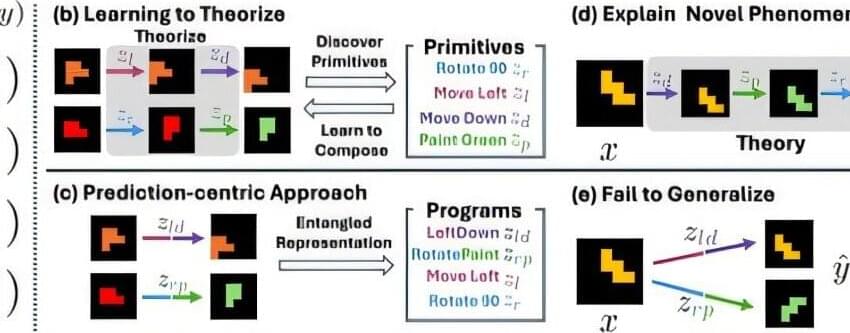
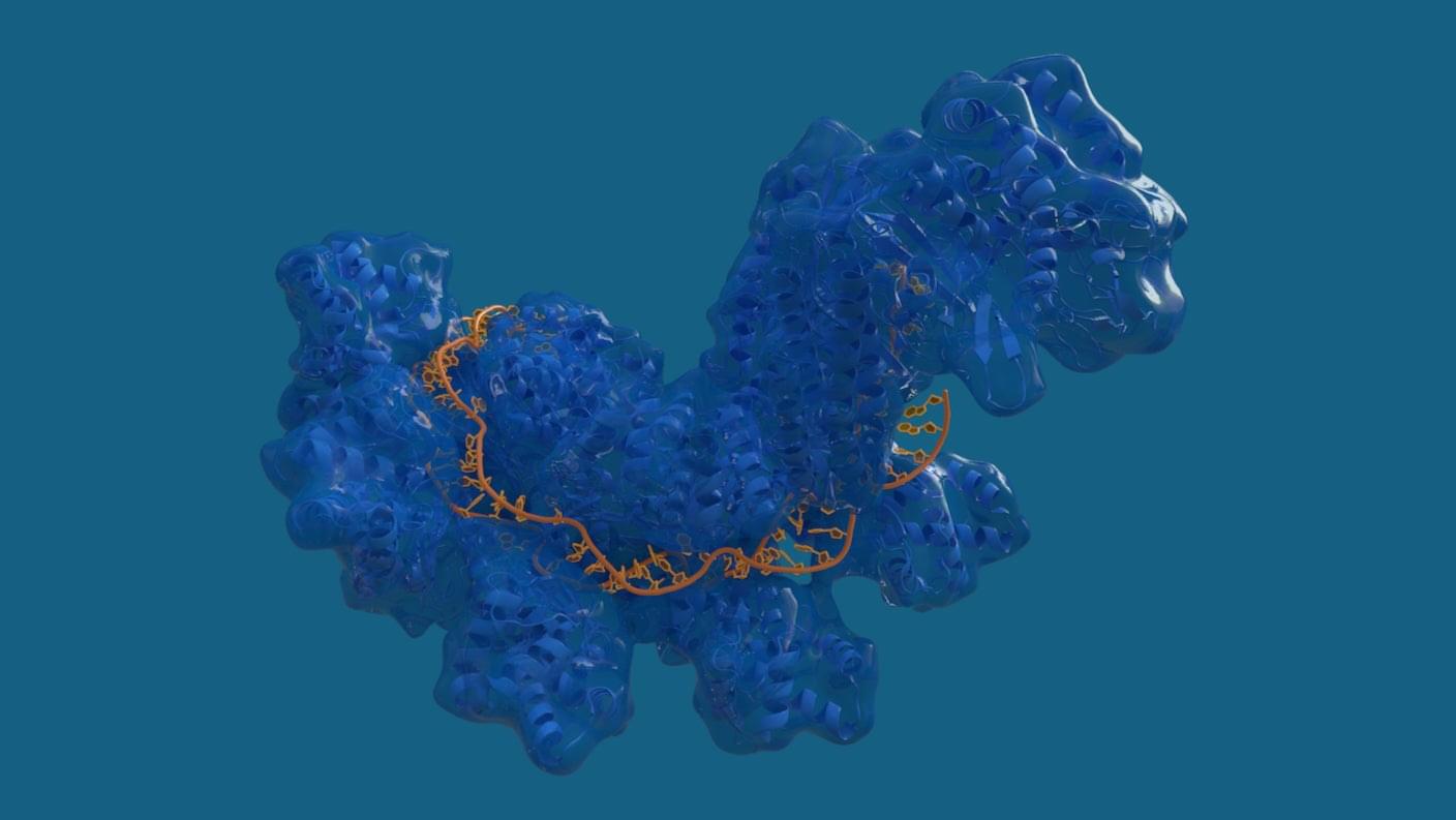
Every encounter between a T cell and a potential target—especially when that target is a developing tumor—begins with a rapid series of molecular decisions. Within seconds, the immune cell must determine whether to launch an attack or stand down. T cells are so potent, so potentially devastating, that misreading the situation can cause serious tissue injury.
But cancer cells come equipped with a bag of tricks that allows them to disarm these powerful warriors of the immune system. All of these activities, whether mediated by T cells or their targets, occur at split-second speed and unfold at the point of cell-to-cell contact.
Now, scientists have identified tiny nanoscale contact points where those decisions are made, revealing how activation and inhibitory signals are integrated at the first moments of a cell-to-cell encounter.
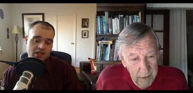
In February 2011, I spent an hour on Skype asking one of the most influential computer scientists alive whether we could still steer the technologies we were building.
James Martin said yes.
He had earned the right to that answer. Computerworld ranked him fourth among the 25 people who most shaped computer science. The Sunday Times called him Britain’s leading futurist. He wrote 104 textbooks, picked up a Pulitzer nomination, collected honorary doctorates from six continents, then gave away more than $100 million to found the Oxford Martin School so 30 institutes could work on the hardest problems of the century.
So when he told me accelerating technology is controllable, he was not being naive. He was being deliberate. Control, in his telling, was never a technical property of the machines. It was a civilizational choice, and he thought this century was the narrow window in which we get to make it.
We talked about exponential growth in genetics, robotics, nanotech and #AI. We talked about The Meaning of the 21st Century and the project he was working on then, the Transformation of Humankind. He was not selling optimism. He was assigning homework.
Fifteen years later, the claim in the title is a lot harder to defend than it was when he made it. Or maybe that is precisely his point, and we are the ones who failed the assignment.


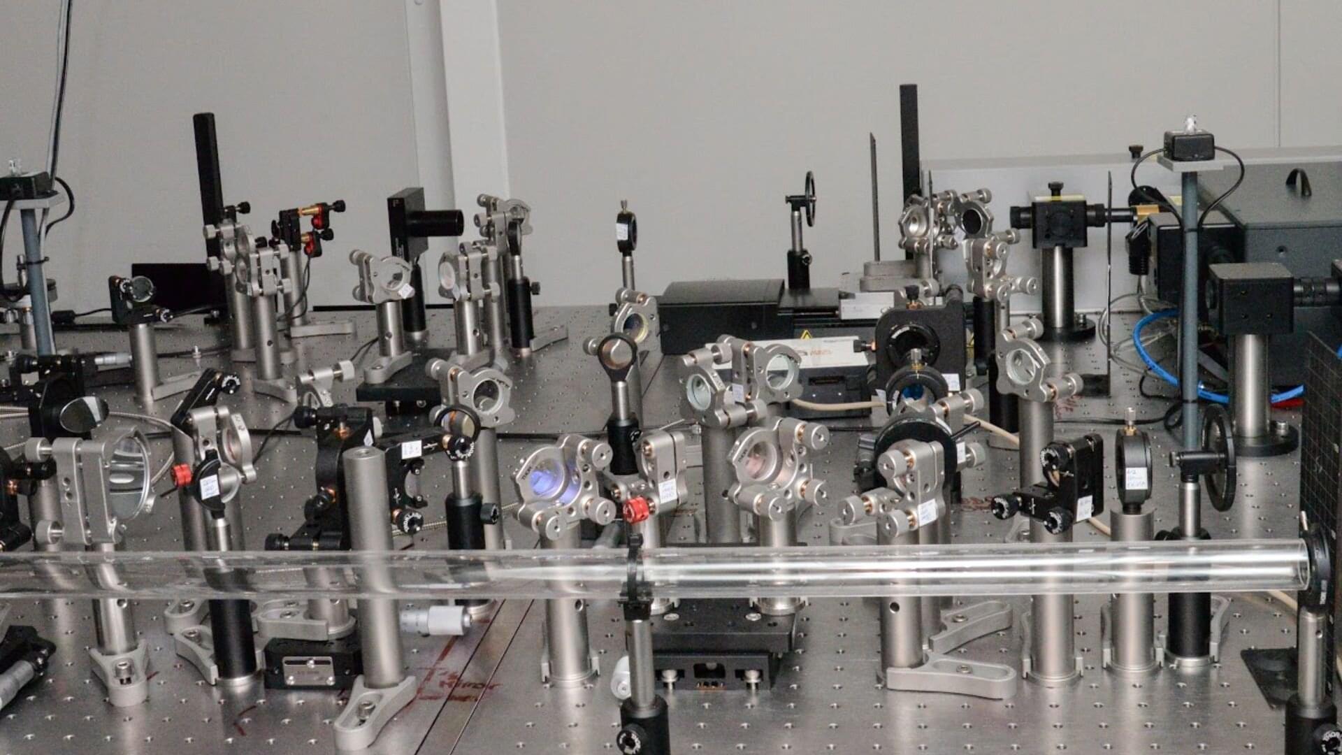
A ring of optical fiber can be made to host phenomena that originated in the realm of high-energy physics.
In non-Abelian gauge theories, particles interact with each other; operation order matters; and a so-called gauge symmetry ensures the invariance of physical laws under local transformations via fields mediated by photons, gluons, and the W and Z bosons. Such theories describe the strong and weak forces; provide the mathematical backbone of the standard model; and underpin efforts to understand the early Universe, quantum gravity, and exotic topological phases of matter. For decades, subjecting these theories to direct experimental scrutiny often meant resorting to enormous particle accelerators. Recently, an experimental approach known as photonic synthetic dimensions has offered a tabletop alternative. Applications of the approach have already realized a non-Abelian magnetic field [1]. Now Shu Yang of the University of Hong Kong and his colleagues have synthesized the corresponding non-Abelian electric field [2].
A new study published in Nature Physics reports the first direct experimental evidence of a Floquet topological state, a novel light-induced phase of matter that, until now, has existed only on paper and in simulations. Topological insulators can conduct electricity along their surface while remaining insulating throughout their bulk. Physicists have spent years developing Floquet engineering, a technique that uses intense, rapidly oscillating light fields to temporarily reshape a material’s electronic structure.
Combining the two ideas seemed like a natural next step: use light to coax an otherwise ordinary material into behaving like a topological insulator on demand. A scheme for realizing such a “Floquet topological insulator” in a semiconductor was proposed in 2011, but pinning down the effect experimentally proved elusive. The predicted state would be short-lived, easy to mistake for other light-matter effects and difficult to disentangle from a material’s ordinary electronic behavior.
Now, researchers have closed that gap using tin telluride (SnTe), a semiconductor that sits close to a topological phase transition. Using femtosecond laser pulses, the team captured direct evidence of the transition.