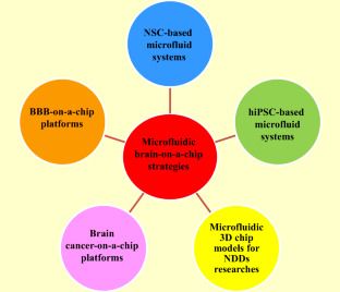
Neurodegenerative diseases (NDDs) include more than 600 types of nervous system disorders in humans that impact tens of millions of people worldwide. Estimates by the World Health Organization (WHO) suggest NDDs will increase by nearly 50% by 2030. Hence, development of advanced models for research on NDDs is needed to explore new therapeutic strategies and explore the pathogenesis of these disorders. Different approaches have been deployed in order to investigate nervous system disorders, including two-and three-dimensional (2D and 3D) cell cultures and animal models. However, these models have limitations, such as lacking cellular tension, fluid shear stress, and compression analysis; thus, studying the biochemical effects of therapeutic molecules on the biophysiological interactions of cells, tissues, and organs is problematic. The microfluidic “organ-on-a-chip” is an inexpensive and rapid analytical technology to create an effective tool for manipulation, monitoring, and assessment of cells, and investigating drug discovery, which enables the culture of various cells in a small amount of fluid (10−9 to 10−18 L). Thus, these chips have the ability to overcome the mentioned restrictions of 2D and 3D cell cultures, as well as animal models. Stem cells (SCs), particularly neural stem cells (NSCs), induced pluripotent stem cells (iPSCs), and embryonic stem cells (ESCs) have the capability to give rise to various neural system cells. Hence, microfluidic organ-on-a-chip and SCs can be used as potential research tools to study the treatment of central nervous system (CNS) and peripheral nervous system (PNS) disorders. Accordingly, in the present review, we discuss the latest progress in microfluidic brain-on-a-chip as a powerful and advanced technology that can be used in basic studies to investigate normal and abnormal functions of the nervous system.


