Toggle light / dark theme

Get the latest international news and world events from around the world.

Log in for authorized contributors

3D microscopy reveals how a tick-borne virus reshapes human cells to replicate

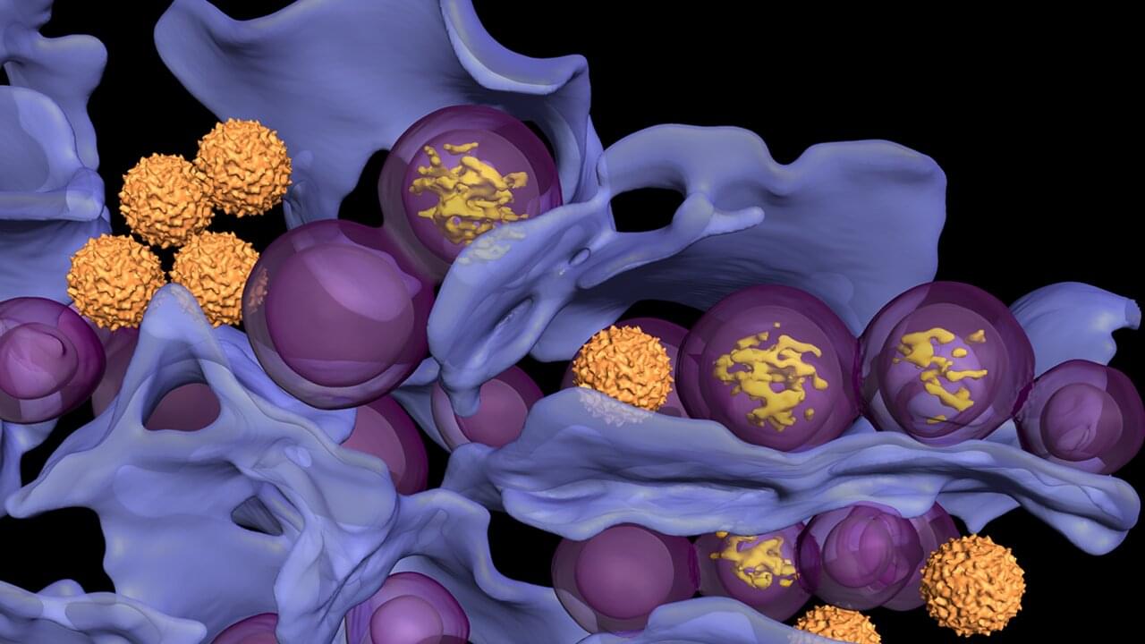
Researchers at Umeå University show how tick-borne viruses remodel human cells into virus factories, using an advanced microscopy method. The findings provide new insight into how the virus replicates and matures, knowledge that may become important for future treatments against TBE. The study is published in Nature Communications.

“When we saw the three-dimensional images for the first time, we immediately realized how much new information we could gain about the virus’s replication,” says Lars-Anders Carlson, professor at the Department of Medical Chemistry and Biophysics at Umeå University, who led the study.

One of the most dangerous viral diseases spread in Europe is tick-borne encephalitis. A bite from an infected tick can transmit the TBE virus to humans and cause severe inflammation of the brain. Using electron microscopy, researchers at Umeå University have now discovered how tick-borne viruses reshape infected human cells and turn them into virus factories.

What this AI epitope library means for vaccines, immunotherapy and biosensors

A new tool makes it possible to screen millions of tiny protein fragments and select those that can be recognized by the immune system. The CIC biomaGUNE Center for Cooperative Research in Biomaterials has developed epiGPTope, a system that uses machine learning to generate and classify epitopes, in collaboration with the company Multiverse Computing.

The immune system is triggered by the presence of viruses or bacteria. When the antibodies produced recognize the epitopes, a small part of these viruses or bacteria, they launch an attack strategy. These epitopes are small fragments of protein recognized by antibodies or by immune cell receptors. So discovering new epitope sequences that target specific antibodies is essential for the development of diagnostic tools, immunotherapies and vaccines.

CIC biomaGUNE’s Biomolecular Nanotechnology laboratory, led by the Ikerbasque Research Professor Aitziber L. Cortajarena, is creating a library or database of hundreds of thousands of synthetic epitopes using this AI-based technique. The work is published in the journal ACS Synthetic Biology.

Metamaterial chains learn new shapes by sharing data hinge to hinge

In a new Nature Physics publication, University of Amsterdam researchers introduce human-made materials that spring to life. These ‘metamaterials’ don’t just learn to change shape, but can autonomously adapt their shape-changing strategy, perform reflex actions and move around like living systems do.

Normal materials have fixed, predetermined responses when a force is applied to them, whereas robots have pre-programmed behaviors. In stark contrast, living materials such as cells and brainless organisms can adapt extremely well to changing conditions. Inspired by nature, the research team created synthetic materials—metamaterials—that learn and adapt without a central “brain.”

The worm-like metamaterials progressively learn how to change shape by being trained on examples. They can forget old shapes and learn new ones, or learn and remember multiple shapes at once and toggle between these shapes. This allows them to perform advanced tasks such as grabbing an object or moving around (locomotion).

A layered approach sharpens brain signals in optical imaging

Near-infrared spectroscopy, or fNIRS, offers a way to monitor brain activity without surgery or radiation by tracking changes in blood flow and oxygenation. Light sources placed on the scalp send near-infrared light into the head, and detectors measure the light that scatters back. Because this light must pass through the scalp and skull before reaching the brain, the measured signal always includes a mix of superficial and cerebral contributions. Separating those signals has long been a central challenge for fNIRS researchers.

In a study published in Biophotonics Discovery, researchers from the Tufts University Diffuse Optical Imaging of Tissue Laboratory show that combining a specific source–detector geometry with a simple, anatomically informed tissue model can substantially improve how fNIRS data are interpreted.

By accounting for how light travels through layered head structures, the approach makes it possible to better isolate brain-specific signals without relying on complex imaging systems or subject-specific MRI scans.

Quantum computing without interruptions

Mid-circuit measurements are one of the biggest practical hurdles in quantum error correction on encoded qubits. Researchers in Innsbruck and Aachen have now proposed and experimentally demonstrated that a universal fault-tolerant quantum algorithm can be executed without such measurements. Using a trapped-ion quantum processor, the team successfully ran Grover’s quantum search algorithm on three logical qubits.

A key bottleneck in today’s leading approaches to quantum error correction is the need to repeatedly pause and measure the quantum processor mid-computation, a process that is slow, technically demanding, and itself a significant source of errors.

Now, a joint team from the University of Innsbruck, RWTH Aachen University, Forschungszentrum Jülich and spin-off Alpine Quantum Technologies (AQT) has demonstrated fault-tolerant quantum computation without any such interruptions.

/* */