How does Alzheimer’s target specific brain cells? Researchers are building a multiscale model of the hippocampus to map neuronal loss and cognitive decline.
New research has linked levels of vitamin D in midlife with toxic tangles of tau protein that accumulate in the brains of those with Alzheimer’s disease.
A statistical analysis of blood samples and brain scans from 793 adults showed that the more vitamin D in someone’s system in middle age, the lower the amount of tau protein tangles they tended to have years later.
The finding comes from an international team of researchers, and while it doesn’t prove direct cause and effect, it suggests an association that’s worth looking at.
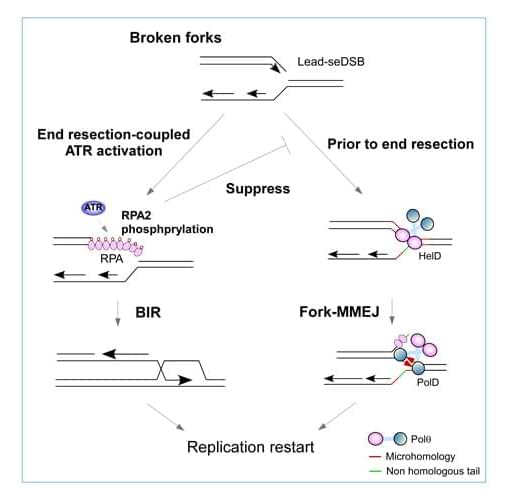
LA JOLLA, CA— A cancer drug target already being investigated in clinical trials turns out to be doing something even more consequential than researchers realized. Scientists at Scripps Research have discovered that the enzyme Pol theta (Polθ) drives a DNA repair mechanism directly at broken replication forks—one of the most frequent forms of DNA damage in cancer cells. The findings, published in Molecular Cell on March 16, 2026, help explain how tumors survive relentless replication stress and clarify why Pol theta inhibitors may be an effective strategy to selectively target cancer.
“We’ve uncovered a whole new dimension of how cancer cells cope with DNA damage at replication forks,” says Xiaohua Wu, professor at Scripps Research and senior author of the study.
Every time a cell divides, it must make an exact copy of its entire genome, a process carried out by molecular machinery that unzips the DNA double helix and reads each strand to build a new one. The point where this unzipping and copying actively happens is called a replication fork. But when this replication machinery encounters damage, forks can stall or collapse, leaving behind dangerous one-ended DNA breaks that are particularly difficult to repair and, if left unresolved, can kill the cell. This is particularly true in cancer cells, where replication stress is constant.
New in JBC press|
asbmbJBC.
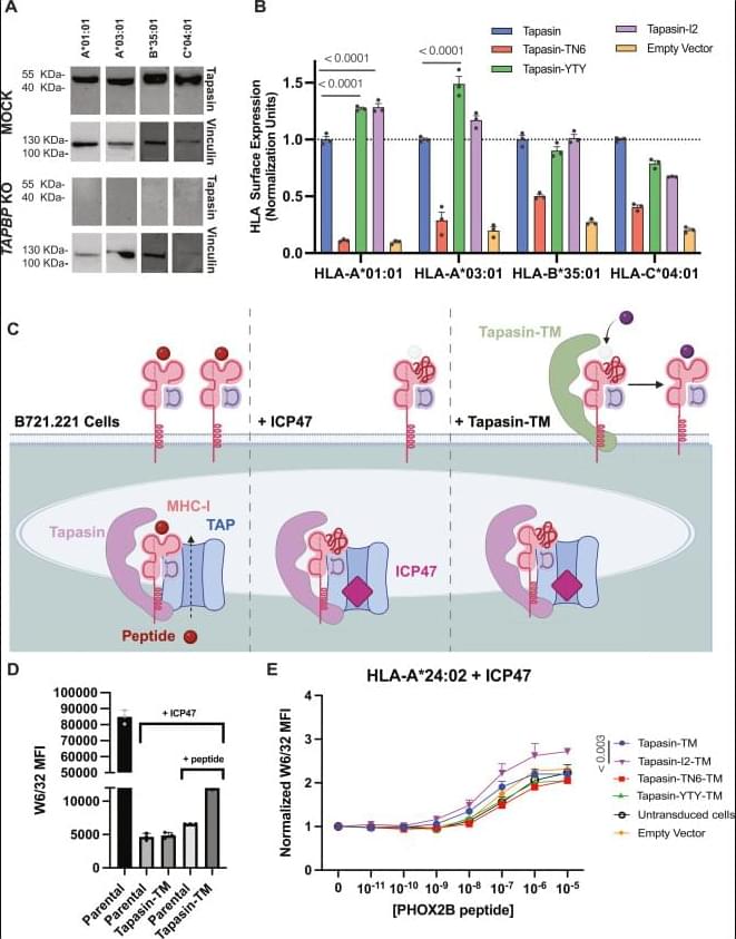
Human leukocyte antigen (HLA) proteins are extremely polymorphic, with different allotypes exhibiting a wide range of dependencies on the chaperone tapasin for peptide loading, expression, and stability at the cell surface. Given its central role in antigen processing, tapasin is frequently downregulated across viral infections and cancers, impairing antigen presentation and hindering the identification of therapeutically relevant peptide antigens. We hypothesized that elucidating the mutational tolerance of tapasin surfaces which mediate interactions with polymorphic HLA residues can provide a means for fine-tuning its chaperoning function and reveal mechanistic epitopes that underlie its function.
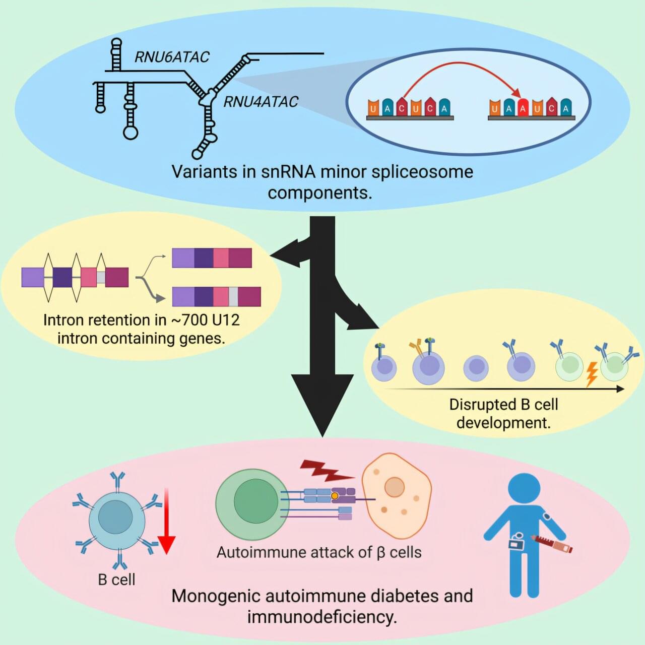
Scientists have found new genetic causes for diabetes in babies—in a part of the genome that has historically been overlooked in genetic studies. Until recently, most research has investigated causes of disease in “coding” genes—those that produce proteins. Now, academics at the University of Exeter and their international collaborators have found that DNA changes in two genes that instead make functional RNA molecules are a cause of diabetes. RNA plays various roles in the body, including regulating genes and influencing how genetic information is “read” and interpreted.
The study is titled “Bi-allelic variants in the non-protein-coding minor spliceosome components RNU6ATAC and RNU4ATAC cause syndromic monogenic autoimmune diabetes,” and was published in the American Journal of Human Genetics.
The team used genome sequencing, a method that reads all the letters in a person’s DNA. They found that changes in two genes called RNU4ATAC and RNU6ATAC were the cause of autoimmune neonatal diabetes in 19 children.

Among patients worldwide with suspected CoronaryArteryDisease, median radiation doses exceeded guideline thresholds (9 mSv) for 21% undergoing nuclear cardiology studies and 44% undergoing CCTA, with dose differences up to 500% across regions.
The findings suggest considerable opportunities to improve cardiac imaging quality and safety through harmonized protocols and technology upgrades.
Question How does radiation dose from cardiac diagnostic testing vary worldwide?
Findings In this cross-sectional study in 101 countries including 19 302 patients, radiation doses varied significantly between imaging tests and among patients receiving the same tests across centers, regions, and country income strata. This was especially pronounced for coronary computed tomography angiography, for which median dose in low-and lower-middle–income countries was more than 280% of that in high-income countries and median dose in Africa was more than 500% of that in Western Europe.
Meaning Current radiation doses for cardiac testing exceeded 9 mSv for 21% of patients undergoing nuclear cardiology studies and for 44% undergoing coronary computed tomography angiography, identifying critical needs for training, standardized protocols, and updated equipment to reduce radiation worldwide.

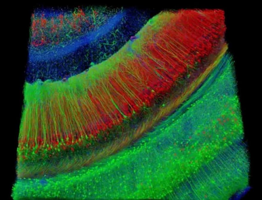
A new Yale School of Medicine (YSM) study has uncovered surprising new details about how our eyes process what we see. When we look at something, our visual system breaks down different aspects of the scene—such as color, contrast, and motion—and processes those components separately. It’s called parallel visual processing and it’s what allows our brains to work out what we’re seeing so quickly.
This separation of information starts in the retina, and scientists have thought that separation is maintained as the information travels through the visual system. But in a study published in Neuron, researchers have found that information channels are more integrated than previously thought. This may help cells process weak visual signals, such as low-light conditions, the researchers say.
“We found that while different channels can deliver their own features, they’re also interconnected by underlying electrical circuitry,” says Yao Xue, Ph.D., a postdoctoral fellow in the department of ophthalmology and visual science at YSM and the study’s first author.