Toggle light / dark theme

Handwriting vs. typing: 30 brain studies reveal which is better for your brain

From the article:

“A review of brain imaging studies found that handwriting activates a broader network of neural pathways than typing or tapping, engaging fine motor skills, memory encoding, and deeper cognitive processing simultaneously. The physical act of forming letters on paper recruits brain regions that digital input simply doesn’t reach. Studies suggest that the pen engages what some researchers describe as a “symphony of neural pathways,” connecting motor control to thought formation in ways that keyboards and touchscreens may bypass.”

Scientists Find Protein Inside The Body That Reverses Brain Aging

Cyclin D-binding myb-like transcription factor 1 or DMTF1a key protein in the brain can help to regenerate neural stem cells and improve aging-associated memory decline. NUS scientists found that this protein’s levels are repressed in the “aged” neural stem cells, Health & Wellness News, Health and Me

Tubulin prevents toxic protein clumps in the brain, fighting back against neurodegeneration

Researchers at Baylor College of Medicine have discovered a potential new strategy to fight back against Alzheimer’s and Parkinson’s diseases, conditions that are linked to the toxic accumulation of Tau and alpha synuclein protein clumps in the brain. The team reports in Nature Communications that tubulin, the building block of microtubules, the cell’s internal ‘railway tracks, can stop Tau and alpha synuclein from forming toxic clumps and instead steer them into their normal, healthy roles.

“Tau and alpha synuclein are well known for their roles in neurodegenerative diseases like Alzheimer’s and Parkinson’s. In these conditions, these proteins can misfold, stick together and form harmful aggregates that damage neurons and contribute to memory loss, movement problems and other symptoms,” said first author Dr. Lathan Lucas, postdoctoral associate of biochemistry and molecular pharmacology in Dr. Allan Ferreon’s lab.

“But Tau and alpha synuclein also fulfill essential functions in healthy neurons—they help maintain cell structure and support communication by interacting with tubulin and contributing to microtubule assembly and stabilization.”

Tubulin Cytoskeleton in Neurodegenerative Diseases–not Only Primary Tubulinopathies

Neurodegenerative diseases represent a large group of disorders characterized by gradual loss of neurons and functions of the central nervous systems. Their course is usually severe, leading to high morbidity and subsequent inability of patients to independent functioning. Vast majority of neurodegenerative diseases is currently untreatable, and only some symptomatic drugs are available which efficacy is usually very limited. To develop novel therapies for this group of diseases, it is crucial to understand their pathogenesis and to recognize factors which can influence the disease course. One of cellular structures which dysfunction appears to be relatively poorly understood in the light of neurodegenerative diseases is tubulin cytoskeleton.

Mitochondrial Ca2+ efflux controls neuronal metabolism and long-term memory across species

Mitochondrial calcium regulates neuronal metabolism and memory.

Brain metabolism is important for long-term memories (LTMs) and various brain functions, Although it is well known that impairing neuronal metabolism limits brain performance, it is not clear if expanding the metabolic capacity of neurons boosts brain function.

In this study, the authors demonstrate that increasing mitochondrial metabolism can enhance LTM formation in flies and mice.

The authors increase mitochondrial Ca2+ by knocking down the mitochondrial Ca2+ exporter Letm1 and demonstrate over-activation of mitochondrial metabolism in neurons of central memory circuits, leading to improved LTM storage. sciencenewshighlights Science Mission https://sciencemission.com/Mitochondrial-Ca2-efflux


Boosting mitochondrial metabolism in neurons in central memory circuits by enhancing Ca2+ retention in the mitochondrial matrix is shown to improve long-term memory formation in flies and mice.

Degenerating Tanycytes Disrupt Tau Removal, Shaping Alzheimer’s Progression

“Tanycytes, whose cell bodies line the walls and floor of the third ventricle and extend long, slim processes that terminate in ‘endfeet’ that contact these fenestrated capillaries,” act as a shuttle between the CSF and the blood, the authors wrote. The new study suggests they also act as a kind of molecular “exit ramp,” moving tau out of the CSF and into the bloodstream for disposal. When these cells become fragmented, that clearance system falters. Tau, which should be ferried away, instead lingers—much like traffic backing up when a major off‑ramp closes—allowing toxic protein species to accumulate.

“Our findings reveal a previously underappreciated, disease‑relevant role for tanycytes in neurodegeneration,” said corresponding author Vincent Prévot, PhD, of INSERM. “Focusing on tanycyte health could be a way to improve tau clearance and limit disease progression.”

Using rodent and cellular models, the researchers showed that tanycytes take up tau from the CSF and release it into pituitary portal capillaries, enabling its entry into the systemic circulation, according to the authors. When the team blocked vesicular transport in tanycytes, tau clearance from CSF to blood slowed dramatically, and tau pathology intensified. As the authors wrote, “Blocking tanycytic vesicular transport blunts CSF‑to‑blood tau efflux and potentiates tau pathology.”

Can a wealthy family change the course of a deadly brain disease?

A wealthy family fighting its own disease boosted research on a little-studied brain protein, progranulin. Can it spur new dementia treatments?


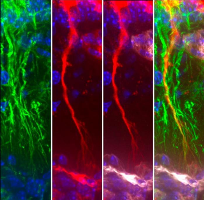
Bluefield investigators, and eventually drug companies, saw something compelling about FTD-GRN, the form of the condition Alice had. In other genetic neurodegenerative disorders, such as familial Alzheimer’s and Huntington disease, mutations spark the production of toxic proteins, generating complex cascades of pathology. But the culprit mutations driving FTD-GRN block progranulin production, leaving carriers with less than half as much of the protein as noncarriers. Many dementia researchers came to describe FTD-GRN as a “low-hanging fruit” among neurodegenerative diseases, using words such as “intuitive” and “tractable” to characterize its biology. The solution seemed obvious: A treatment just needed to raise progranulin levels in the brain.

Fueled in part by that confidence, six clinical trials have been launched to test progranulin-boosting therapies in FTD-GRN. Companies also hope the anti-inflammatory properties of a progranulin-boosting agent could help in Parkinson’s disease, Alzheimer’s, amyotrophic lateral sclerosis (ALS), and FTD caused by other mutations or without a known genetic cause.

All has not gone according to plan, however. In October 2025, a landmark phase 3 clinical trial of a progranulin-boosting drug in people with FTD-GRN did not keep their disease from progressing. In February, a small trial of a gene therapy delivering a healthy copy of GRN to the brain was halted, also for lack of effect.

Researchers detect complex emotions by combining multiple optical signals

Researchers have developed a new way to recognize human emotions by combining fiber-based physiological signals with thermal images of the face. The portable emotional recognition system could eventually be used to support at-home mental health monitoring, improve driver safety and make technology more responsive to human emotions.

“Unlike many existing approaches, our method does not rely on facial expressions, which can be consciously controlled or exaggerated,” said research team leader Rui Min from Beijing Normal University at Zhuhai (BNU Zhuhai) in China.

“Instead, it focuses on natural bodily responses that occur automatically. By combining multiple types of optical sensing and higher-level physiological interpretation, our system can more accurately capture emotion than those using a single type of input.”

New study shows how sickle cell affects brain function

Sickle cell disease is often thought of solely as a blood disorder, but new research from the Wood Neuro Research Group provides measurable evidence that it can reshape how brain networks function. Previous neuroimaging studies have relied on functional connectivity to show that adults with sickle cell disease may experience changes in how brain networks communicate among one another, potentially compensating for reduced oxygen delivery. However, this method is limited in determining the directionality or influence between networks.

“Red blood cells that carry oxygen to the brain are altered by the disease, resulting in reduced oxygen delivery to all regions of the brain and long-term changes in how it functions,” outlined Nahom Mossazghi, biomedical engineering Ph.D. student and the study’s first author. “The brain actively recruits other regions to help process information, which we do not see in people without the disease.”

The study, published in Human Brain Mapping, used MRI and advanced analytical tools originally developed in economics to examine how different brain networks influence one another. Instead of functional connectivity, effective connectivity was used to address a gap in the field and interpret how specific networks support one another in response to the disease-related changes.

Temporal lobe epilepsy: A new strategy to correct abnormal electrical activity

Many patients suffer from epilepsy that cannot be controlled by current medications. Surgical removal of epileptogenic brain regions is effective in only about half of cases, and not all patients are eligible for the procedure. For these individuals, therapeutic options remain severely limited. Researchers from the Paris Brain Institute and the Institut du Fer à Moulin in Paris have now taken an important step forward: they have identified two molecules capable of reducing seizure frequency by targeting a mechanism that has so far received little attention. Their findings are published in Proceedings of the National Academy of Sciences.

For the brain to function normally, it must continuously regulate its electrical activity. One of the key mechanisms involved is GABAergic signaling, a natural inhibitory system that controls neuronal activity and prevents the electrical bursts that characterize epileptic seizures. This braking system depends on a delicate balance: the concentration of chloride inside neurons.

An ion transporter known as KCC2 is responsible for removing excess chloride from nerve cells. When it functions poorly—as observed in many neurological disorders, including mesial temporal lobe epilepsy, the most common form of focal epilepsy in adults—chloride accumulates inside neurons. As a result, GABAergic signals, instead of inhibiting neuronal activity, can paradoxically excite it.

/* */