Toggle light / dark theme

Scientists find a mechanism showing how exercise protects the brain

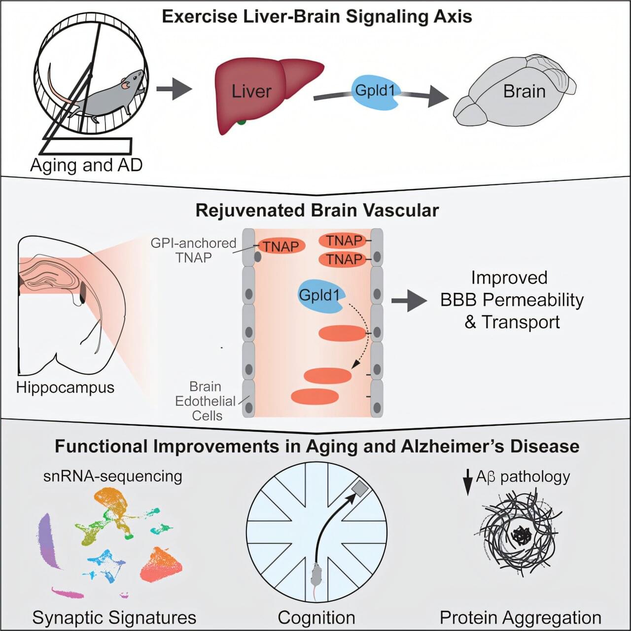
Researchers at UC San Francisco have discovered a mechanism that could explain how exercise improves cognition by shoring up the brain’s protective barrier. With age, the network of blood vessels—called the blood–brain barrier—gets leaky, letting harmful compounds enter the brain. This causes inflammation, which is associated with cognitive decline and is seen in conditions like Alzheimer’s disease. The research is published in the journal Cell.

Six years ago, the team identified a brain-rejuvenating enzyme called GPLD1 that mice produced in their livers when they exercised. But they couldn’t understand how it worked, because it cannot get into the brain.

The new study answers that question. Researchers discovered that GPLD1 was working through another protein called TNAP. As the mice age, the cells that form the blood-brain barrier accumulate TNAP, which makes it leaky. But when mice exercise, their livers produce GPLD1. It travels to the vessels that surround the brain and trims TNAP off the cells.

Pop-up-style 3D electrode array captures organoid-wide brain rhythms in real time

A team led by Northwestern University and Shirley Ryan AbilityLab scientists have developed a new technology that can eavesdrop on the hidden electrical dialogues unfolding inside miniature, lab-grown human brain-like tissues. Known as human neural organoids—and sometimes called “mini brains”—these millimeter-sized structures are powerful models of brain development and disease. But until now, scientists could only record and stimulate activity from a small fraction of their neurons—missing network-wide dynamics that give rise to coordinated rhythms, information processing and the complex patterns of activity that define brain function.

For the first time, the new technology overcomes that stubborn limitation. The soft, three-dimensional (3D) electronic framework wraps around an organoid like a breathable, high-tech mesh. Rather than sampling select regions, it delivers near-complete, shape-conforming coverage with hundreds of miniaturized electrodes. That dense, three-dimensional interfacing enables scientists to map and manipulate neural activity across almost the entire organoid.

By moving from localized probing to true whole-network mapping, the work brings organoid research closer to capturing how real human brains develop, function and even fail.

How ADHD Stimulants

In a large US-based brain imaging study, researchers found that these drugs do not primarily affect attention networks, but instead act on systems linked to arousal, sleep, and motivation.

The puzzle of ADHD stimulants

Prescription stimulants such as methylphenidate and amphetamines are among the most used psychoactive drugs in children and adolescents with ADHD, where they remain a first-line treatment. Estimates for receiving a prescription for ADHD medication among diagnosed children vary from 38–81%. Despite their widespread use, there is still disagreement about how these drugs work in the brain.

Sjögren Syndrome Candidate Autoantigen AQP5 Triggers AQP4 CNS Autoimmunity Through Self-Antigen Mimicry

Tumor-immune-neural circuit in cancer cachexia.

The mechanisms involved in cancer-mediated cachexia and anorexia are not well understood.

The researchers in this study delineate an interplay among tumor cells, immune cells, and the nervous system that drives cancer cachexia and anorexia.

The authors show thay loss of GDF15 protects against appetite loss, muscle wasting, and fat loss in pancreatic, lung, and skin cancers.

Disrupting this feedforward loop with GDF15-neutralizing antibody, anti-CSF1R antibody, or Rearranged during Transfection (RET) inhibitor alleviates cachexia and anorexia across cancer models. sciencenewshighlights ScienceMission https://sciencemission.com/Tumor-immune-neural-circuit


Shi et al. delineate an interplay among tumor cells, immune cells, and the nervous system that drives cancer cachexia and anorexia. Specifically, tumor-derived CSF1 induces macrophage GDF15, which signals through the GFRAL-RET neural axis to enhance β-adrenergic activity and systemic wasting. Disrupting this feedforward loop alleviates cachexia across cancer models.

A single inhalation of vapor from dried toad secretion containing 5-methoxy-N

See my Comment below for a link to the original article in NATURE.


5-methoxy-N, N-dimethyltryptamine (hereinafter referred to as 5-MeO-DMT) is a psychedelic substance found in the secretion from the parotoid glands of the Bufo alvarius toad. Inhalation of vapor from toad secretion containing 5-MeO-DMT has become popular in naturalistic settings as a treatment of mental health problems or as a means for spiritual exploration. However, knowledge of the effects of 5-MeO-DMT in humans is limited.

Developmental ‘switch’ in brain may shape lifelong obesity risk

Researchers at UT Southwestern Medical Center have discovered that a crucial developmental process in the brain’s hypothalamus may influence how susceptible individuals are to obesity. Their preclinical findings, published in Neuron, show that a transcription factor called Otp acts as a molecular “switch” that directs immature hypothalamic neurons toward either appetite-suppressing or appetite-stimulating fates—their ultimate identities as specialized cells. The researchers found that disrupting this switch alters feeding behavior and protects mice from diet-induced obesity.

“These findings show that early developmental decisions in the hypothalamus have a long-lasting impact on energy balance,” said senior author Chen Liu, Ph.D., Associate Professor of Internal Medicine and Neuroscience and an Investigator in the Peter O’Donnell Jr. Brain Institute at UT Southwestern.

“By uncovering this fate-switching program, we can begin to understand how the brain establishes lifelong metabolic set points.”

The enigma of reflex eating epilepsy: A cohort study of 50 patients with insights from multimodal evaluation

“EE is a disabling form of reflex epilepsy with heterogeneous clinical, EEG and neuroimaging features, which are not necessarily substrate-specific. Findings from our study point to the presence of a wide epileptogenic network prominently involving perisylvian regions. Treatment outcomes in drug-refractory EE remain suboptimal, and further studies are needed for a better understanding and management of this complex entity.”

Read this original article from Epileptic Disorders at doi.org/10.1002/epd2.70132.


Objectives To evaluate the clinical, electroencephalographic (EEG), neuroimaging characteristics, and treatment outcomes of patients diagnosed with eating epilepsy (EE). Methods This retrospective study was conducted at a tertiary care epilepsy referral center in India. Patients diagnosed with EE between 2002 and 2025, with at least one EEG and magnetic resonance imaging (MRI) available for review, were consecutively included. Clinical data and multimodal evaluation findings including video EEG, brain MRI, positron emission tomography-MRI (PET-MRI), and magnetoencephalography (MEG) were systematically collected using a structured proforma. Seizure outcomes and treatment strategies were subsequently analyzed.

/* */